


Crystal Structure of the Prokaryotic Crotonase
Bruning, J.B., Delgado, E., Ghosh, S., Sacchettini, J.C.To be published.
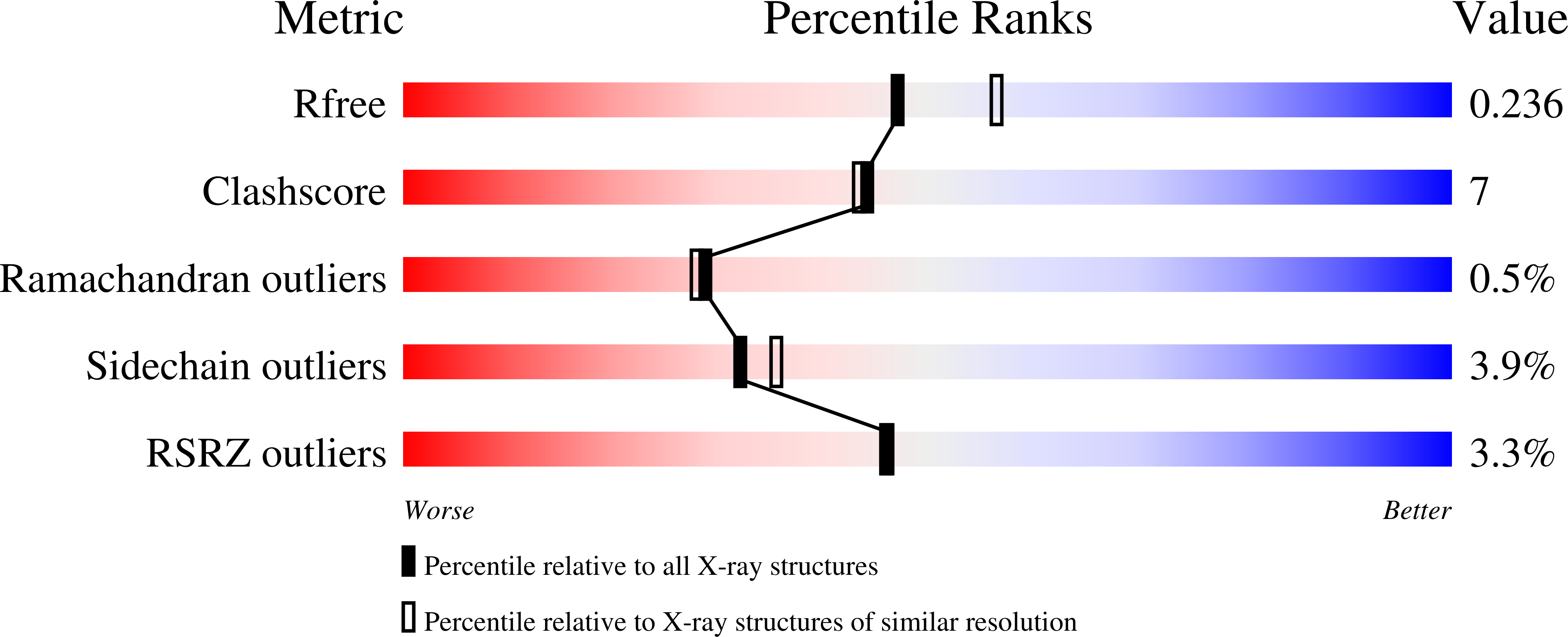
Experimental Data Snapshot
wwPDB Validation 3D Report Full Report
Entity ID: 1 | |||||
|---|---|---|---|---|---|
| Molecule | Chains | Sequence Length | Organism | Details | Image |
| e enoyl-CoA hydratase echA8 | 257 | Mycobacterium tuberculosis | Mutation(s): 0 Gene Names: echA8, MT1100, MTV017.23c, Rv1070c EC: 4.2.1.17 |  | |
UniProt | |||||
Find proteins for P9WNN9 (Mycobacterium tuberculosis (strain ATCC 25618 / H37Rv)) Explore P9WNN9 Go to UniProtKB: P9WNN9 | |||||
Entity Groups | |||||
| Sequence Clusters | 30% Identity50% Identity70% Identity90% Identity95% Identity100% Identity | ||||
| UniProt Group | P9WNN9 | ||||
Sequence AnnotationsExpand | |||||
| |||||
| Ligands 1 Unique | |||||
|---|---|---|---|---|---|
| ID | Chains | Name / Formula / InChI Key | 2D Diagram | 3D Interactions | |
| SO4 Query on SO4 | D [auth A], E [auth B] | SULFATE ION O4 S QAOWNCQODCNURD-UHFFFAOYSA-L |  | ||
| Length ( Å ) | Angle ( ˚ ) |
|---|---|
| a = 133.796 | α = 90 |
| b = 133.734 | β = 90 |
| c = 103.45 | γ = 90 |
| Software Name | Purpose |
|---|---|
| SCALEPACK | data scaling |
| PHASER | phasing |
| PHENIX | refinement |
| PDB_EXTRACT | data extraction |
| HKL-2000 | data collection |
| DENZO | data reduction |
RCSB PDB (citation) is hosted by
RCSB PDB is a member of the

