3PS9

Crystal structure of MnmC from E. coli


Experimental Data Snapshot

  • Method: X-RAY DIFFRACTION
  • Resolution: 2.54 Å
  • R-Value Free: 0.247 
  • R-Value Work: 0.182 
  • R-Value Observed: 0.185 

wwPDB Validation   3D Report Full Report


Ligand Structure Quality Assessment 


This is version 1.3 of the entry. See complete history


Literature

Structural basis for hypermodification of the wobble uridine in tRNA by bifunctional enzyme MnmC.

Kim, J.Almo, S.C.

(2013) BMC Struct Biol 13: 5-5

  • DOI: https://doi.org/10.1186/1472-6807-13-5
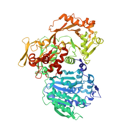
  • Primary Citation of Related Structures:  
    3PS9, 3PVC, 3SGL

  • PubMed Abstract: 

    Methylaminomethyl modification of uridine or 2-thiouridine (mnm5U34 or mnm5s2U34) at the wobble position of tRNAs specific for glutamate, lysine and arginine are observed in Escherichia coli and allow for specific recognition of codons ending in A or G. In the biosynthetic pathway responsible for this post-transcriptional modification, the bifunctional enzyme MnmC catalyzes the conversion of its hypermodified substrate carboxymethylaminomethyl uridine (cmnm5U34) to mnm5U34. MnmC catalyzes the flavin adenine dinucleotide (FAD)-dependent oxidative cleavage of carboxymethyl group from cmnm5U34 via an imine intermediate to generate aminomethyl uridine (nm5U34), which is subsequently methylated by S-adenosyl-L-methionine (SAM) to yield methylaminomethyl uridine (mnm5U34).


  • Organizational Affiliation

    Albert Einstein College of Medicine, 1300 Morris Park Avenue, Bronx, New York 10461, USA. jukim@aecom.yu.edu


Macromolecules
Find similar proteins by:  (by identity cutoff)  |  3D Structure
Entity ID: 1
MoleculeChains Sequence LengthOrganismDetailsImage
tRNA 5-methylaminomethyl-2-thiouridine biosynthesis bifunctional protein mnmC676Escherichia coli BL21Mutation(s): 0 
Gene Names: B21_02209mnmCyfcK
EC: 2.1.1.61
Entity Groups  
Sequence Clusters30% Identity50% Identity70% Identity90% Identity95% Identity100% Identity
Sequence Annotations
Expand
  • Reference Sequence
Experimental Data & Validation

Experimental Data

  • Method: X-RAY DIFFRACTION
  • Resolution: 2.54 Å
  • R-Value Free: 0.247 
  • R-Value Work: 0.182 
  • R-Value Observed: 0.185 
  • Space Group: P 41 21 2
Unit Cell:
Length ( Å )Angle ( ˚ )
a = 100.063α = 90
b = 100.063β = 90
c = 159.19γ = 90
Software Package:
Software NamePurpose
HKL-2000data collection
MOLREPphasing
REFMACrefinement
HKL-2000data reduction
HKL-2000data scaling

Structure Validation

View Full Validation Report


Ligand Structure Quality Assessment 


Entry History 

Deposition Data

Revision History  (Full details and data files)

  • Version 1.0: 2010-12-22
    Type: Initial release
  • Version 1.1: 2011-07-13
    Changes: Source and taxonomy, Version format compliance
  • Version 1.2: 2013-08-21
    Changes: Database references
  • Version 1.3: 2024-02-21
    Changes: Data collection, Database references, Derived calculations