3PGQ

Crystal Structure of the Carboxyltransferase Domain of S. cerevisiae Acetyl CoA Carboxylase in Complex with Pinoxaden


Experimental Data Snapshot

  • Method: X-RAY DIFFRACTION
  • Resolution: 2.80 Å
  • R-Value Free: 0.239 
  • R-Value Work: 0.197 
  • R-Value Observed: 0.199 

wwPDB Validation   3D Report Full Report


Ligand Structure Quality Assessment 


This is version 1.3 of the entry. See complete history


Literature

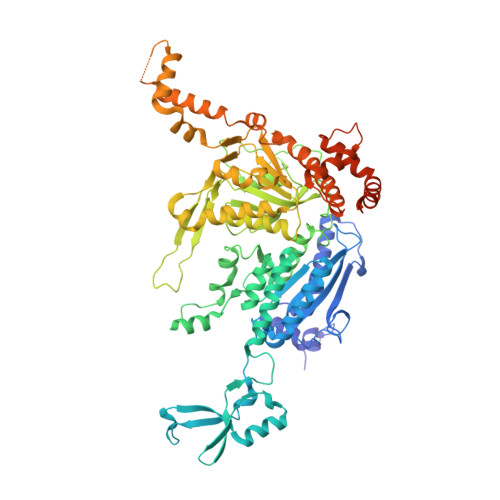
Mechanism for the inhibition of the carboxyltransferase domain of acetyl-coenzyme A carboxylase by pinoxaden.

Yu, L.P.Kim, Y.S.Tong, L.

(2010) Proc Natl Acad Sci U S A 107: 22072-22077

  • DOI: https://doi.org/10.1073/pnas.1012039107
  • Primary Citation of Related Structures:  
    3PGQ

  • PubMed Abstract: 

    Acetyl-CoA carboxylases (ACCs) are crucial metabolic enzymes and have been targeted for drug development against obesity, diabetes, and other diseases. The carboxyltransferase (CT) domain of this enzyme is the site of action for three different classes of herbicides, as represented by haloxyfop, tepraloxydim, and pinoxaden. Our earlier studies have demonstrated that haloxyfop and tepraloxydim bind in the CT active site at the interface of its dimer. However, the two compounds probe distinct regions of the dimer interface, sharing primarily only two common anchoring points of interaction with the enzyme. We report here the crystal structure of the CT domain of yeast ACC in complex with pinoxaden at 2.8-Å resolution. Despite their chemical diversity, pinoxaden has a similar binding mode as tepraloxydim and requires a small conformational change in the dimer interface for binding. Crystal structures of the CT domain in complex with all three classes of herbicides confirm the importance of the two anchoring points for herbicide binding. The structures also provide a foundation for understanding the molecular basis of the herbicide resistance mutations and cross resistance among the herbicides, as well as for the design and development of new inhibitors against plant and human ACCs.


  • Organizational Affiliation

    Department of Biological Sciences, Columbia University, New York, NY 10027, USA.


Macromolecules
Find similar proteins by:  (by identity cutoff)  |  3D Structure
Entity ID: 1
MoleculeChains Sequence LengthOrganismDetailsImage
Acetyl-CoA carboxylase
A, B, C
764Saccharomyces cerevisiaeMutation(s): 0 
Gene Names: FAS3ACC1YNR016CN3175
EC: 6.4.1.2
UniProt
Find proteins for Q00955 (Saccharomyces cerevisiae (strain ATCC 204508 / S288c))
Explore Q00955 
Go to UniProtKB:  Q00955
Entity Groups  
Sequence Clusters30% Identity50% Identity70% Identity90% Identity95% Identity100% Identity
UniProt GroupQ00955
Sequence Annotations
Expand
  • Reference Sequence
Small Molecules
Ligands 1 Unique
IDChains Name / Formula / InChI Key2D Diagram3D Interactions
GY3
Query on GY3

Download Ideal Coordinates CCD File 
D [auth A],
E [auth B],
F [auth B]
8-(2-ethenyl-6-ethyl-4-methylphenyl)tetrahydro-7H-pyrazolo[1,2-d][1,4,5]oxadiazepine-7,9(8H)-dione
C18 H22 N2 O3
CYYDMVKNZPACLF-UHFFFAOYSA-N
Experimental Data & Validation

Experimental Data

  • Method: X-RAY DIFFRACTION
  • Resolution: 2.80 Å
  • R-Value Free: 0.239 
  • R-Value Work: 0.197 
  • R-Value Observed: 0.199 
  • Space Group: C 1 2 1
Unit Cell:
Length ( Å )Angle ( ˚ )
a = 247.18α = 90
b = 123.415β = 94.29
c = 145.711γ = 90
Software Package:
Software NamePurpose
DENZOdata reduction
SCALEPACKdata scaling
COMOphasing
REFMACrefinement
PDB_EXTRACTdata extraction
HKL-2000data collection
HKL-2000data reduction
HKL-2000data scaling

Structure Validation

View Full Validation Report


Ligand Structure Quality Assessment 


Entry History 

Deposition Data

Revision History  (Full details and data files)

  • Version 1.0: 2010-12-22
    Type: Initial release
  • Version 1.1: 2011-07-13
    Changes: Version format compliance
  • Version 1.2: 2017-11-08
    Changes: Refinement description
  • Version 1.3: 2024-02-21
    Changes: Data collection, Database references, Derived calculations