3PCM

STRUCTURE OF PROTOCATECHUATE 3,4-DIOXYGENASE COMPLEXED WITH 6-HYDROXYNICOTINIC ACID N-OXIDE AND CYANIDE


Experimental Data Snapshot

  • Method: X-RAY DIFFRACTION
  • Resolution: 2.25 Å
  • R-Value Work: 0.180 

wwPDB Validation   3D Report Full Report


This is version 2.0 of the entry. See complete history


Literature

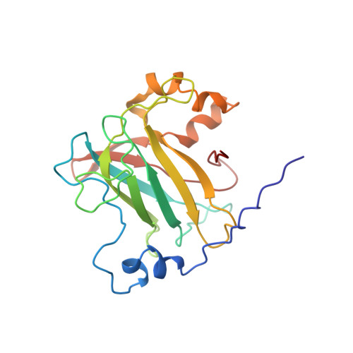
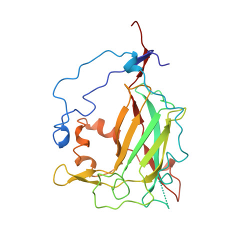
Crystal structures of substrate and substrate analog complexes of protocatechuate 3,4-dioxygenase: endogenous Fe3+ ligand displacement in response to substrate binding.

Orville, A.M.Lipscomb, J.D.Ohlendorf, D.H.

(1997) Biochemistry 36: 10052-10066

  • DOI: https://doi.org/10.1021/bi970469f
  • Primary Citation of Related Structures:  
    3PCA, 3PCJ, 3PCK, 3PCL, 3PCM

  • PubMed Abstract: 

    Protocatechuate 3,4-dioxygenase (3,4-PCD) utilizes a ferric ion to catalyze the aromatic ring cleavage of 3,4-dihydroxybenzoate (PCA) by incorporation of both atoms of dioxygen to yield beta-carboxy-cis, cis-muconate. The crystal structures of the anaerobic 3,4-PCD.PCA complex, aerobic complexes with two heterocyclic PCA analogs, 2-hydroxyisonicotinic acid N-oxide (INO) and 6-hydroxynicotinic acid N-oxide (NNO), and ternary complexes of 3,4-PCD.INO.CN and 3,4-PCD. NNO.CN have been determined at 2.1-2.2 A resolution and refined to R-factors between 0.165 and 0.184. PCA, INO, and NNO form very similar, asymmetrically chelated complexes with the active site Fe3+ that result in dissociation of the endogenous axial tyrosinate Fe3+ ligand, Tyr447 (147beta). After its release from the iron, Tyr447 is stabilized by hydrogen bonding to Tyr16 (16alpha) and Asp413 (113beta) and forms the top of a small cavity adjacent to the C3-C4 bond of PCA. The equatorial Fe3+ coordination site within this cavity is unoccupied in the anaerobic 3,4-PCD.PCA complex but coordinates a solvent molecule in the 3,4-PCD.INO and 3,4-PCD.NNO complexes and CN- in the 3,4-PCD.INO.CN and 3,4-PCD.NNO.CN complexes. This shows that an O2 analog can occupy the cavity and suggests that electrophilic O2 attack on PCA is initiated from this site. Both the dissociation of the endogenous Tyr447 and the expansion of the iron coordination sphere are novel features of the 3,4-PCD. substrate complex which appear to play essential roles in the activation of substrate for O2 attack. Together, the structures presented here and in the preceding paper [Orville, A. M., Elango, N. , Lipscomb, J. D., & Ohlendorf, D. H. (1997) Biochemistry 36, 10039-10051] provide atomic models for several steps in the reaction cycle of 3,4-PCD and related Fe3+-containing dioxygenases.


  • Organizational Affiliation

    Department of Biochemistry, Medical School, and Center for Metals in Biocatalysis, University of Minnesota, Minneapolis, Minnesota 55455-0347, USA.


Macromolecules
Find similar proteins by:  (by identity cutoff)  |  3D Structure
Entity ID: 1
MoleculeChains Sequence LengthOrganismDetailsImage
PROTOCATECHUATE 3,4-DIOXYGENASE200Pseudomonas putidaMutation(s): 0 
EC: 1.13.11.3
UniProt
Find proteins for P00436 (Pseudomonas putida)
Explore P00436 
Go to UniProtKB:  P00436
Entity Groups  
Sequence Clusters30% Identity50% Identity70% Identity90% Identity95% Identity100% Identity
UniProt GroupP00436
Sequence Annotations
Expand
  • Reference Sequence
Find similar proteins by:  (by identity cutoff)  |  3D Structure
Entity ID: 2
MoleculeChains Sequence LengthOrganismDetailsImage
PROTOCATECHUATE 3,4-DIOXYGENASE238Pseudomonas putidaMutation(s): 0 
EC: 1.13.11.3
UniProt
Find proteins for P00437 (Pseudomonas putida)
Explore P00437 
Go to UniProtKB:  P00437
Entity Groups  
Sequence Clusters30% Identity50% Identity70% Identity90% Identity95% Identity100% Identity
UniProt GroupP00437
Sequence Annotations
Expand
  • Reference Sequence
Small Molecules
Ligands 3 Unique
IDChains Name / Formula / InChI Key2D Diagram3D Interactions
NNO
Query on NNO

Download Ideal Coordinates CCD File 
AA [auth Q]
DA [auth R]
O [auth M]
R [auth N]
U [auth O]
AA [auth Q],
DA [auth R],
O [auth M],
R [auth N],
U [auth O],
X [auth P]
6-HYDROXYISONICOTINIC ACID N-OXIDE
C6 H5 N O4
PQTDJLRPWRPPLV-UHFFFAOYSA-N
FE
Query on FE

Download Ideal Coordinates CCD File 
CA [auth R]
N [auth M]
Q [auth N]
T [auth O]
W [auth P]
CA [auth R],
N [auth M],
Q [auth N],
T [auth O],
W [auth P],
Z [auth Q]
FE (III) ION
Fe
VTLYFUHAOXGGBS-UHFFFAOYSA-N
CYN
Query on CYN

Download Ideal Coordinates CCD File 
BA [auth R]
M
P [auth N]
S [auth O]
V [auth P]
BA [auth R],
M,
P [auth N],
S [auth O],
V [auth P],
Y [auth Q]
CYANIDE ION
C N
XFXPMWWXUTWYJX-UHFFFAOYSA-N
Experimental Data & Validation

Experimental Data

  • Method: X-RAY DIFFRACTION
  • Resolution: 2.25 Å
  • R-Value Work: 0.180 
  • Space Group: I 1 2 1
Unit Cell:
Length ( Å )Angle ( ˚ )
a = 196.1α = 90
b = 127.3β = 97.7
c = 134.5γ = 90
Software Package:
Software NamePurpose
PROLSQrefinement
XENGENdata reduction
XENGENdata scaling

Structure Validation

View Full Validation Report


Entry History 

Deposition Data

Revision History  (Full details and data files)

  • Version 1.0: 1998-01-21
    Type: Initial release
  • Version 1.1: 2008-03-25
    Changes: Version format compliance
  • Version 1.2: 2011-07-13
    Changes: Advisory, Derived calculations, Version format compliance
  • Version 2.0: 2023-09-27
    Type: Remediation
    Changes: Advisory, Atomic model, Data collection, Database references, Derived calculations, Other, Refinement description