3P3Y

Crystal structure of neurofascin homophilic adhesion complex in space group p6522


Experimental Data Snapshot

  • Method: X-RAY DIFFRACTION
  • Resolution: 2.60 Å
  • R-Value Free: 0.288 
  • R-Value Work: 0.258 
  • R-Value Observed: 0.258 

wwPDB Validation   3D Report Full Report


Ligand Structure Quality Assessment 


This is version 1.2 of the entry. See complete history


Literature

Homophilic adhesion mechanism of neurofascin, a member of the l1 family of neural cell adhesion molecules.

Liu, H.Focia, P.J.He, X.

(2011) J Biol Chem 286: 797-805

  • DOI: https://doi.org/10.1074/jbc.M110.180281
  • Primary Citation of Related Structures:  
    3P3Y, 3P40

  • PubMed Abstract: 

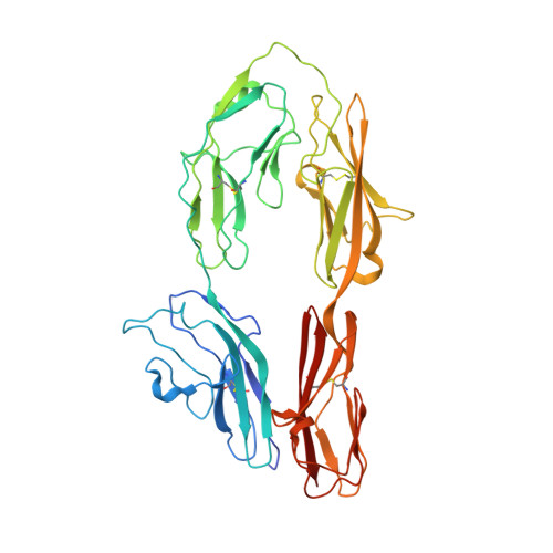
    The L1 family neural cell adhesion molecules play key roles in specifying the formation and remodeling of the neural network, but their homophilic interaction that mediates adhesion is not well understood. We report two crystal structures of a dimeric form of the headpiece of neurofascin, an L1 family member. The four N-terminal Ig-like domains of neurofascin form a horseshoe shape, akin to several other immunoglobulin superfamily cell adhesion molecules such as hemolin, axonin, and Dscam. The neurofascin dimer, captured in two crystal forms with independent packing patterns, reveals a pair of horseshoes in trans-synaptic adhesion mode. The adhesion interaction is mediated mostly by the second Ig-like domain, which features an intermolecular β-sheet formed by the joining of two individual GFC β-sheets and a large but loosely packed hydrophobic cluster. Mutagenesis combined with gel filtration assays suggested that the side chain hydrogen bonds at the intermolecular β-sheet are essential for the homophilic interaction and that the residues at the hydrophobic cluster play supplementary roles. Our structures reveal a conserved homophilic adhesion mode for the L1 family and also shed light on how the pathological mutations of L1 affect its structure and function.


  • Organizational Affiliation

    Department of Molecular Pharmacology and Biological Chemistry, Northwestern University Feinberg School of Medicine, Chicago, Illinois 60611, USA.


Macromolecules
Find similar proteins by:  (by identity cutoff)  |  3D Structure
Entity ID: 1
MoleculeChains Sequence LengthOrganismDetailsImage
Neurofascin404Homo sapiensMutation(s): 0 
Gene Names: NFASCKIAA0756
UniProt & NIH Common Fund Data Resources
Find proteins for O94856 (Homo sapiens)
Explore O94856 
Go to UniProtKB:  O94856
PHAROS:  O94856
GTEx:  ENSG00000163531 
Entity Groups  
Sequence Clusters30% Identity50% Identity70% Identity90% Identity95% Identity100% Identity
UniProt GroupO94856
Sequence Annotations
Expand
  • Reference Sequence
Small Molecules
Ligands 1 Unique
IDChains Name / Formula / InChI Key2D Diagram3D Interactions
NAG
Query on NAG

Download Ideal Coordinates CCD File 
B [auth A]2-acetamido-2-deoxy-beta-D-glucopyranose
C8 H15 N O6
OVRNDRQMDRJTHS-FMDGEEDCSA-N
Experimental Data & Validation

Experimental Data

  • Method: X-RAY DIFFRACTION
  • Resolution: 2.60 Å
  • R-Value Free: 0.288 
  • R-Value Work: 0.258 
  • R-Value Observed: 0.258 
  • Space Group: P 65 2 2
Unit Cell:
Length ( Å )Angle ( ˚ )
a = 171.425α = 90
b = 171.425β = 90
c = 87.792γ = 120
Software Package:
Software NamePurpose
HKL-2000data collection
SOLVEphasing
CNSrefinement
HKL-2000data reduction
HKL-2000data scaling

Structure Validation

View Full Validation Report


Ligand Structure Quality Assessment 


Entry History 

Deposition Data

  • Released Date: 2010-11-03 
  • Deposition Author(s): Liu, H., He, X.

Revision History  (Full details and data files)

  • Version 1.0: 2010-11-03
    Type: Initial release
  • Version 1.1: 2011-07-13
    Changes: Version format compliance
  • Version 1.2: 2020-07-29
    Type: Remediation
    Reason: Carbohydrate remediation
    Changes: Data collection, Derived calculations, Structure summary