3P19

Improved NADPH-dependent Blue Fluorescent Protein


Experimental Data Snapshot

  • Method: X-RAY DIFFRACTION
  • Resolution: 2.05 Å
  • R-Value Free: 0.239 
  • R-Value Work: 0.186 
  • R-Value Observed: 0.189 

wwPDB Validation   3D Report Full Report


Ligand Structure Quality Assessment 


This is version 1.2 of the entry. See complete history


Literature

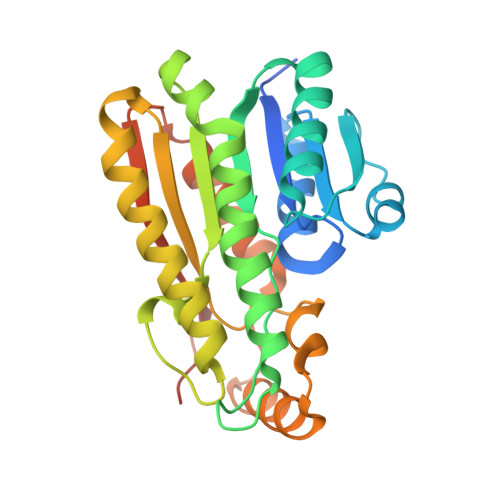
Structure of a NADPH-dependent blue fluorescent protein revealed the unique role of Gly176 on the fluorescence enhancement.

Kao, T.H.Chen, Y.Pai, C.H.Chang, M.C.Wang, A.H.J.

(2011) J Struct Biol 174: 485-493

  • DOI: https://doi.org/10.1016/j.jsb.2011.02.010
  • Primary Citation of Related Structures:  
    3P19

  • PubMed Abstract: 

    A NADPH-dependent blue fluorescent protein from Vibrio vulnificus CKM-1 (BFPvv) emits blue fluorescence under UV-exposure. Previously, the BFPvvD7 mutant generated by directed evolution displayed a fourfold enhancement in fluorescent intensity. Herein, a further increase in fluorescence in the new BFPvvD8 mutant, with three additional mutations from BFPvvD7, was made. To understand the underlying mechanism of the increased fluorescent intensity of BFPvv, we solved the BFPvvD8-NADPH complex structure. Accompanied with lifetime detection, we proposed that the enhanced intensity is related to the conformational change caused by a glycine residue (Gly176) mutated to other non-glycine residues at a turn close to the NADPH binding site. We also observed the Förster resonance energy transfer (FRET) from our BFPvvD8 to each of the GFP-like fluorescent proteins, mTFP1 and EGFP, joined by an eight-residue linker between the N-terminal of BFPvvD8 and the C-terminal of GFPs. Taken together, with the newly solved BFPvvD8 structure, our results not only provide new considerations within the rational-based protein engineering of this NADPH-dependent BFP, but also suggest that BFPvvD8 could be a potential candidate in FRET-based biosensor techniques.


  • Organizational Affiliation

    Institute of Biological Chemistry, Academia Sinica, Taipei, Taiwan.


Macromolecules
Find similar proteins by:  (by identity cutoff)  |  3D Structure
Entity ID: 1
MoleculeChains Sequence LengthOrganismDetailsImage
Putative blue fluorescent protein
A, B, C, D
266Vibrio vulnificusMutation(s): 0 
UniProt
Find proteins for Q9F172 (Vibrio vulnificus)
Explore Q9F172 
Go to UniProtKB:  Q9F172
Entity Groups  
Sequence Clusters30% Identity50% Identity70% Identity90% Identity95% Identity100% Identity
UniProt GroupQ9F172
Sequence Annotations
Expand
  • Reference Sequence
Experimental Data & Validation

Experimental Data

  • Method: X-RAY DIFFRACTION
  • Resolution: 2.05 Å
  • R-Value Free: 0.239 
  • R-Value Work: 0.186 
  • R-Value Observed: 0.189 
  • Space Group: P 43
Unit Cell:
Length ( Å )Angle ( ˚ )
a = 64.403α = 90
b = 64.403β = 90
c = 262.588γ = 90
Software Package:
Software NamePurpose
REFMACrefinement
PDB_EXTRACTdata extraction
HKL-2000data collection
HKL-2000data reduction
HKL-2000data scaling
PHASERphasing

Structure Validation

View Full Validation Report


Ligand Structure Quality Assessment 


Entry History 

Deposition Data

Revision History  (Full details and data files)

  • Version 1.0: 2011-07-20
    Type: Initial release
  • Version 1.1: 2013-11-20
    Changes: Non-polymer description
  • Version 1.2: 2023-11-01
    Changes: Data collection, Database references, Refinement description