Structural and functional analysis of phytotoxin toxoflavin-degrading enzyme
Jung, W.S., Lee, J., Kim, M.I., Ma, J., Nagamatsu, T., Goo, E., Kim, H., Hwang, I., Han, J., Rhee, S.(2011) PLoS One 6: e22443-e22443
- PubMed: 21799856
- DOI: https://doi.org/10.1371/journal.pone.0022443
- Primary Citation of Related Structures:
3OUL, 3OUM - PubMed Abstract:
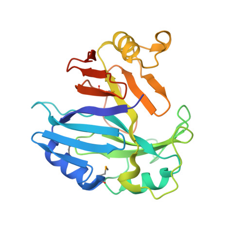
Pathogenic bacteria synthesize and secrete toxic low molecular weight compounds as virulence factors. These microbial toxins play essential roles in the pathogenicity of bacteria in various hosts, and are emerging as targets for antivirulence strategies. Toxoflavin, a phytotoxin produced by Burkholderia glumae BGR1, has been known to be the key factor in rice grain rot and wilt in many field crops. Recently, toxoflavin-degrading enzyme (TxDE) was identified from Paenibacillus polymyxa JH2, thereby providing a possible antivirulence strategy for toxoflavin-mediated plant diseases. Here, we report the crystal structure of TxDE in the substrate-free form and in complex with toxoflavin, along with the results of a functional analysis. The overall structure of TxDE is similar to those of the vicinal oxygen chelate superfamily of metalloenzymes, despite the lack of apparent sequence identity. The active site is located at the end of the hydrophobic channel, 9 Å in length, and contains a Mn(II) ion interacting with one histidine residue, two glutamate residues, and three water molecules in an octahedral coordination. In the complex, toxoflavin binds in the hydrophobic active site, specifically the Mn(II)-coordination shell by replacing a ligating water molecule. A functional analysis indicated that TxDE catalyzes the degradation of toxoflavin in a manner dependent on oxygen, Mn(II), and the reducing agent dithiothreitol. These results provide the structural features of TxDE and the early events in catalysis.
Organizational Affiliation:
Department of Agricultural Biotechnology, Seoul National University, Seoul, Korea.