3OU0

re-refined 3CS0


Experimental Data Snapshot

  • Method: X-RAY DIFFRACTION
  • Resolution: 3.00 Å
  • R-Value Free: 0.229 
  • R-Value Work: 0.196 
  • R-Value Observed: 0.197 

wwPDB Validation   3D Report Full Report


This is version 1.3 of the entry. See complete history

Re-refinement Note

This entry reflects an alternative modeling of the original data in: 3CS0


Literature

Covalent Linkage of Distinct Substrate Degrons Controls Assembly and Disassembly of DegP Proteolytic Cages.

Kim, S.Grant, R.A.Sauer, R.T.

(2011) Cell 145: 67-78

  • DOI: https://doi.org/10.1016/j.cell.2011.02.024
  • Primary Citation of Related Structures:  
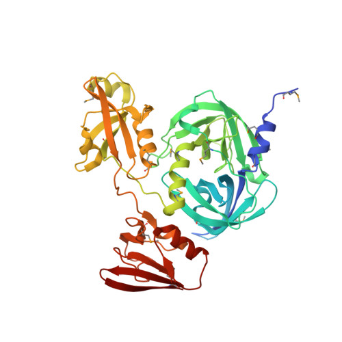
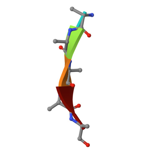
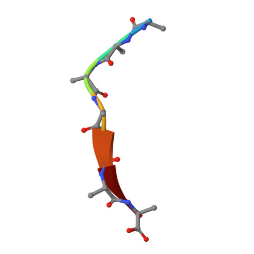
    3OTP, 3OU0

  • PubMed Abstract: 

    Protein quality control requires careful regulation of intracellular proteolysis. For DegP, a periplasmic protease, substrates promote assembly of inactive hexamers into proteolytically active cages with 12, 18, 24, or 30 subunits. Here, we show that sensitive activation and cage assembly require covalent linkage of distinct substrate sequences that affect degradation (degrons). One degron binds the DegP active site, and another degron binds a separate tethering site in PDZ1 in the crystal structure of a substrate-bound DegP dodecamer. FRET experiments demonstrate that active cages assemble rapidly in a reaction that is positively cooperative in substrate concentration, remain stably assembled while uncleaved substrate is present, and dissociate once degradation is complete. Thus, the energy of binding of linked substrate degrons drives assembly of the proteolytic machine responsible for subsequent degradation. Substrate cleavage and depletion results in disassembly, ensuring that DegP is proteolytically active only when sufficient quantities of protein substrates are present.


  • Organizational Affiliation

    Department of Biology, Massachusetts Institute of Technology, Cambridge, MA 02139, USA.


Macromolecules
Find similar proteins by:  (by identity cutoff)  |  3D Structure
Entity ID: 1
MoleculeChains Sequence LengthOrganismDetailsImage
Periplasmic serine endoprotease DegP448Escherichia coliMutation(s): 1 
Gene Names: b0161degPhtrAJW0157ptd
EC: 3.4.21
UniProt
Find proteins for P0C0V0 (Escherichia coli (strain K12))
Explore P0C0V0 
Go to UniProtKB:  P0C0V0
Entity Groups  
Sequence Clusters30% Identity50% Identity70% Identity90% Identity95% Identity100% Identity
UniProt GroupP0C0V0
Sequence Annotations
Expand
  • Reference Sequence

Find similar proteins by:  Sequence   |   3D Structure  

Entity ID: 2
MoleculeChains Sequence LengthOrganismDetailsImage
pentapeptide5N/AMutation(s): 0 
Sequence Annotations
Expand
  • Reference Sequence

Find similar proteins by:  Sequence   |   3D Structure  

Entity ID: 3
MoleculeChains Sequence LengthOrganismDetailsImage
heptapeptide7N/AMutation(s): 0 
Sequence Annotations
Expand
  • Reference Sequence
Small Molecules
Modified Residues  1 Unique
IDChains TypeFormula2D DiagramParent
MSE
Query on MSE
A
L-PEPTIDE LINKINGC5 H11 N O2 SeMET
Experimental Data & Validation

Experimental Data

  • Method: X-RAY DIFFRACTION
  • Resolution: 3.00 Å
  • R-Value Free: 0.229 
  • R-Value Work: 0.196 
  • R-Value Observed: 0.197 
  • Space Group: F 4 3 2
Unit Cell:
Length ( Å )Angle ( ˚ )
a = 253.929α = 90
b = 253.929β = 90
c = 253.929γ = 90
Software Package:
Software NamePurpose
PHENIXrefinement
PDB_EXTRACTdata extraction
ADSCdata collection
XDSdata reduction
XSCALEdata scaling
SHARPphasing

Structure Validation

View Full Validation Report


Entry History 

Deposition Data

Revision History  (Full details and data files)

  • Version 1.0: 2011-01-19
    Type: Initial release
  • Version 1.1: 2011-07-13
    Changes: Version format compliance
  • Version 1.2: 2011-10-12
    Changes: Other
  • Version 1.3: 2012-02-22
    Changes: Other