Subunit dissociation and metal binding by Escherichia coli apo-manganese superoxide dismutase.
Whittaker, M.M., Lerch, T.F., Kirillova, O., Chapman, M.S., Whittaker, J.W.(2011) Arch Biochem Biophys 505: 213-225
- PubMed: 21044611
- DOI: https://doi.org/10.1016/j.abb.2010.10.021
- Primary Citation of Related Structures:
3OT7 - PubMed Abstract:
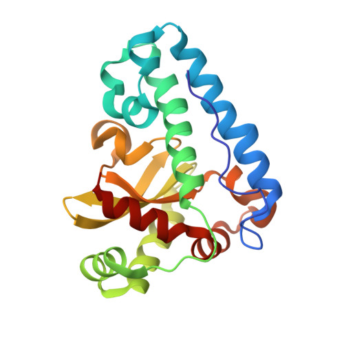
Metal binding by apo-manganese superoxide dismutase (apo-MnSOD) is essential for functional maturation of the enzyme. Previous studies have demonstrated that metal binding by apo-MnSOD is conformationally gated, requiring protein reorganization for the metal to bind. We have now solved the X-ray crystal structure of apo-MnSOD at 1.9Å resolution. The organization of active site residues is independent of the presence of the metal cofactor, demonstrating that protein itself templates the unusual metal coordination geometry. Electrophoretic analysis of mixtures of apo- and (Mn₂)-MnSOD, dye-conjugated protein, or C-terminal Strep-tag II fusion protein reveals a dynamic subunit exchange process associated with cooperative metal binding by the two subunits of the dimeric protein. In contrast, (S126C) (SS) apo-MnSOD, which contains an inter-subunit covalent disulfide-crosslink, exhibits anti-cooperative metal binding. The protein concentration dependence of metal uptake kinetics implies that protein dissociation is involved in metal binding by the wild type apo-protein, although other processes may also contribute to gating metal uptake. Protein concentration dependent small-zone size exclusion chromatography is consistent with apo-MnSOD dimer dissociation at low protein concentration (K(D)=1×10⁻⁵ M). Studies on metal uptake by apo-MnSOD in Escherichia coli cells show that the protein exhibits similar behavior in vivo and in vitro.
Organizational Affiliation:
Institute for Environmental Health, Oregon Health and Science University, Beaverton, 97006-8921, USA.