3ONR

Crystal structure of the calcium chelating immunodominant antigen, calcium dodecin (Rv0379),from Mycobacterium tuberculosis with a novel calcium-binding site


Experimental Data Snapshot

  • Method: X-RAY DIFFRACTION
  • Resolution: 1.80 Å
  • R-Value Free: 0.229 
  • R-Value Work: 0.181 
  • R-Value Observed: 0.183 

wwPDB Validation   3D Report Full Report


This is version 1.2 of the entry. See complete history


Literature

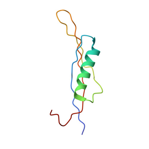
Crystal structure of calcium dodecin (Rv0379), from Mycobacterium tuberculosis with a unique calcium-binding site.

Arockiasamy, A.Aggarwal, A.Savva, C.G.Holzenburg, A.Sacchettini, J.C.

(2011) Protein Sci 20: 827-833

  • DOI: https://doi.org/10.1002/pro.607
  • Primary Citation of Related Structures:  
    3ONR

  • PubMed Abstract: 

    In eukaryotes, calcium-binding proteins play a pivotal role in diverse cellular processes, and recent findings suggest similar roles for bacterial proteins at different stages in their life cycle. Here, we report the crystal structure of calcium dodecin, Rv0379, from Mycobacterium tuberculosis with a dodecameric oligomeric assembly and a unique calcium-binding motif. Structure and sequence analysis were used to identify orthologs of Rv0379 with different ligand-binding specificity.


  • Organizational Affiliation

    Department of Biochemistry and Biophysics, Texas A&M University, College Station, Texas 77843-2128, USA.


Macromolecules
Find similar proteins by:  (by identity cutoff)  |  3D Structure
Entity ID: 1
MoleculeChains Sequence LengthOrganismDetailsImage
PROTEIN TRANSPORT PROTEIN SECE2
A, B, C, D, E
A, B, C, D, E, F, G, H, I, J, K, L
72Mycobacterium tuberculosisMutation(s): 2 
Gene Names: Rv0379secE2
UniProt
Find proteins for Q6MX43 (Mycobacterium tuberculosis (strain ATCC 25618 / H37Rv))
Explore Q6MX43 
Go to UniProtKB:  Q6MX43
Entity Groups  
Sequence Clusters30% Identity50% Identity70% Identity90% Identity95% Identity100% Identity
UniProt GroupQ6MX43
Sequence Annotations
Expand
  • Reference Sequence
Small Molecules
Ligands 3 Unique
IDChains Name / Formula / InChI Key2D Diagram3D Interactions
PT
Query on PT

Download Ideal Coordinates CCD File 
AA [auth I],
T [auth E],
V [auth F]
PLATINUM (II) ION
Pt
HRGDZIGMBDGFTC-UHFFFAOYSA-N
FMT
Query on FMT

Download Ideal Coordinates CCD File 
CA [auth J]
DA [auth K]
EA [auth L]
N [auth A]
O [auth B]
CA [auth J],
DA [auth K],
EA [auth L],
N [auth A],
O [auth B],
P [auth C],
R [auth D],
S [auth E],
U [auth F],
X [auth G],
Y [auth H],
Z [auth I]
FORMIC ACID
C H2 O2
BDAGIHXWWSANSR-UHFFFAOYSA-N
CA
Query on CA

Download Ideal Coordinates CCD File 
BA [auth J],
M [auth A],
Q [auth D],
W [auth G]
CALCIUM ION
Ca
BHPQYMZQTOCNFJ-UHFFFAOYSA-N
Experimental Data & Validation

Experimental Data

  • Method: X-RAY DIFFRACTION
  • Resolution: 1.80 Å
  • R-Value Free: 0.229 
  • R-Value Work: 0.181 
  • R-Value Observed: 0.183 
  • Space Group: P 21 21 21
Unit Cell:
Length ( Å )Angle ( ˚ )
a = 70.193α = 90
b = 78.446β = 90
c = 152.489γ = 90
Software Package:
Software NamePurpose
REFMACrefinement
PDB_EXTRACTdata extraction
HKL-2000data reduction
HKL-2000data scaling
SHARPphasing

Structure Validation

View Full Validation Report


Entry History 

Deposition Data

Revision History  (Full details and data files)

  • Version 1.0: 2011-03-16
    Type: Initial release
  • Version 1.1: 2011-07-13
    Changes: Version format compliance
  • Version 1.2: 2024-02-21
    Changes: Data collection, Database references, Derived calculations