3NVI

Structure of N-terminal truncated Nop56/58 bound with L7Ae and box C/D RNA


Experimental Data Snapshot

  • Method: X-RAY DIFFRACTION
  • Resolution: 2.71 Å
  • R-Value Free: 0.249 
  • R-Value Work: 0.208 
  • R-Value Observed: 0.210 

wwPDB Validation   3D Report Full Report


This is version 1.2 of the entry. See complete history


Literature

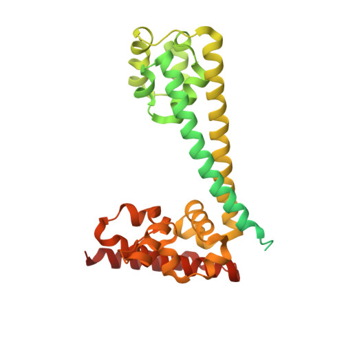
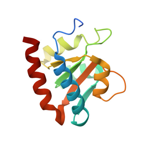
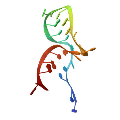
Structural basis for substrate placement by an archaeal box C/D ribonucleoprotein particle.

Xue, S.Wang, R.Yang, F.Terns, R.M.Terns, M.P.Zhang, X.Maxwell, E.S.Li, H.

(2010) Mol Cell 39: 939-949

  • DOI: https://doi.org/10.1016/j.molcel.2010.08.022
  • Primary Citation of Related Structures:  
    3NMU, 3NVI, 3NVK, 3NVM

  • PubMed Abstract: 

    Box C/D small nucleolar and Cajal body ribonucleoprotein particles (sno/scaRNPs) direct site-specific 2'-O-methylation of ribosomal and spliceosomal RNAs and are critical for gene expression. Here we report crystal structures of an archaeal box C/D RNP containing three core proteins (fibrillarin, Nop56/58, and L7Ae) and a half-mer box C/D guide RNA paired with a substrate RNA. The structure reveals a guide-substrate RNA duplex orientation imposed by a composite protein surface and the conserved GAEK motif of Nop56/58. Molecular modeling supports a dual C/D RNP structure that closely mimics that recently visualized by electron microscopy. The substrate-bound dual RNP model predicts an asymmetric protein distribution between the RNP that binds and methylates the substrate RNA. The predicted asymmetric nature of the holoenzyme is consistent with previous biochemical data on RNP assembly and provides a simple solution for accommodating base-pairing between the C/D guide RNA and large ribosomal and spliceosomal substrate RNAs.


  • Organizational Affiliation

    Institute of Molecular Biophysics, Florida State University, Tallahassee, FL 32306, USA.


Macromolecules

Find similar proteins by:  (by identity cutoff)  |  3D Structure
Entity ID: 1
MoleculeChains Sequence LengthOrganismDetailsImage
NOP5/NOP56 related protein
A, C
379Pyrococcus furiosus DSM 3638Mutation(s): 0 
Gene Names: PF0060
UniProt
Find proteins for Q8U4M1 (Pyrococcus furiosus (strain ATCC 43587 / DSM 3638 / JCM 8422 / Vc1))
Explore Q8U4M1 
Go to UniProtKB:  Q8U4M1
Entity Groups  
Sequence Clusters30% Identity50% Identity70% Identity90% Identity95% Identity100% Identity
UniProt GroupQ8U4M1
Sequence Annotations
Expand
  • Reference Sequence
Find similar proteins by:  (by identity cutoff)  |  3D Structure
Entity ID: 2
MoleculeChains Sequence LengthOrganismDetailsImage
50S ribosomal protein L7Ae
B, D
129Pyrococcus furiosus DSM 3638Mutation(s): 0 
Gene Names: PF1367rpl7ae
UniProt
Find proteins for Q8U160 (Pyrococcus furiosus (strain ATCC 43587 / DSM 3638 / JCM 8422 / Vc1))
Explore Q8U160 
Go to UniProtKB:  Q8U160
Entity Groups  
Sequence Clusters30% Identity50% Identity70% Identity90% Identity95% Identity100% Identity
UniProt GroupQ8U160
Sequence Annotations
Expand
  • Reference Sequence

Find similar nucleic acids by:  Sequence   |   3D Structure  

Entity ID: 3
MoleculeChains LengthOrganismImage
RNA (5'-R(*CP*UP*CP*UP*GP*AP*CP*CP*GP*AP*AP*AP*GP*GP*CP*GP*UP*GP*AP*UP*GP*AP*GP*C)-3')
E, F
24Pyrococcus furiosus DSM 3638
Sequence Annotations
Expand
  • Reference Sequence
Experimental Data & Validation

Experimental Data

  • Method: X-RAY DIFFRACTION
  • Resolution: 2.71 Å
  • R-Value Free: 0.249 
  • R-Value Work: 0.208 
  • R-Value Observed: 0.210 
  • Space Group: P 21 21 21
Unit Cell:
Length ( Å )Angle ( ˚ )
a = 87.216α = 90
b = 91.841β = 90
c = 155.603γ = 90
Software Package:
Software NamePurpose
CBASSdata collection
PHENIXmodel building
PHENIXrefinement
HKL-2000data reduction
HKL-2000data scaling
PHENIXphasing

Structure Validation

View Full Validation Report


Entry History 

Deposition Data

Revision History  (Full details and data files)

  • Version 1.0: 2011-07-20
    Type: Initial release
  • Version 1.1: 2013-09-25
    Changes: Derived calculations
  • Version 1.2: 2024-02-21
    Changes: Data collection, Database references