Molecular basis of vancomycin dependence in VanA-type Staphylococcus aureus VRSA-9.
Meziane-Cherif, D., Saul, F.A., Moubareck, C., Weber, P., Haouz, A., Courvalin, P., Perichon, B.(2010) J Bacteriol 192: 5465-5471
- PubMed: 20729361
- DOI: https://doi.org/10.1128/JB.00613-10
- Primary Citation of Related Structures:
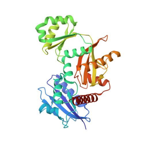
3N8D - PubMed Abstract:
The vancomycin-resistant Staphylococcus aureus VRSA-9 clinical isolate was partially dependent on glycopeptide for growth. The responsible vanA operon had the same organization as that of Tn1546 and was located on a plasmid. The chromosomal D-Ala:D-Ala ligase (ddl) gene had two point mutations that led to Q260K and A283E substitutions, resulting in a 200-fold decrease in enzymatic activity compared to that of the wild-type strain VRSA-6. To gain insight into the mechanism of enzyme impairment, we determined the crystal structure of VRSA-9 Ddl and showed that the A283E mutation induces new ion pair/hydrogen bond interactions, leading to an asymmetric rearrangement of side chains in the dimer interface. The Q260K substitution is located in an exposed external loop and did not induce any significant conformational change. The VRSA-9 strain was susceptible to oxacillin due to synthesis of pentadepsipeptide precursors ending in D-alanyl-D-lactate which are not substrates for the β-lactam-resistant penicillin binding protein PBP2'. Comparison with the partially vancomycin-dependent VRSA-7, whose Ddl is 5-fold less efficient than that of VRSA-9, indicated that the levels of vancomycin dependence and susceptibility to β-lactams correlate with the degree of Ddl impairment. Ddl drug targeting could therefore be an effective strategy against vancomycin-resistant S. aureus.
Organizational Affiliation:
Institut Pasteur, Unité des Agents Antibactériens, CNRS-URA 2185, 25 rue du Docteur Roux, 75724 Paris Cedex 15, France.