3MHH

Structure of the SAGA Ubp8/Sgf11/Sus1/Sgf73 DUB module


Experimental Data Snapshot

  • Method: X-RAY DIFFRACTION
  • Resolution: 2.45 Å
  • R-Value Free: 0.273 
  • R-Value Work: 0.202 
  • R-Value Observed: 0.205 

wwPDB Validation   3D Report Full Report


This is version 1.2 of the entry. See complete history


Literature

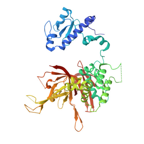
Structural insights into the assembly and function of the SAGA deubiquitinating module.

Samara, N.L.Datta, A.B.Berndsen, C.E.Zhang, X.Yao, T.Cohen, R.E.Wolberger, C.

(2010) Science 328: 1025-1029

  • DOI: https://doi.org/10.1126/science.1190049
  • Primary Citation of Related Structures:  
    3MHH, 3MHS

  • PubMed Abstract: 

    SAGA is a transcriptional coactivator complex that is conserved across eukaryotes and performs multiple functions during transcriptional activation and elongation. One role is deubiquitination of histone H2B, and this activity resides in a distinct subcomplex called the deubiquitinating module (DUBm), which contains the ubiquitin-specific protease Ubp8, bound to Sgf11, Sus1, and Sgf73. The deubiquitinating activity depends on the presence of all four DUBm proteins. We report here the 1.90 angstrom resolution crystal structure of the DUBm bound to ubiquitin aldehyde, as well as the 2.45 angstrom resolution structure of the uncomplexed DUBm. The structure reveals an arrangement of protein domains that gives rise to a highly interconnected complex, which is stabilized by eight structural zinc atoms that are critical for enzymatic activity. The structure suggests a model for how interactions with the other DUBm proteins activate Ubp8 and allows us to speculate about how the DUBm binds to monoubiquitinated histone H2B in nucleosomes.


  • Organizational Affiliation

    Department of Biophysics and Biophysical Chemistry, The Johns Hopkins University School of Medicine, Baltimore, MD 21205, USA.


Macromolecules
Find similar proteins by:  (by identity cutoff)  |  3D Structure
Entity ID: 1
MoleculeChains Sequence LengthOrganismDetailsImage
Ubiquitin carboxyl-terminal hydrolase 8476Saccharomyces cerevisiaeMutation(s): 0 
Gene Names: UBP8YMR223WYM9959.05
EC: 3.1.2.15
UniProt
Find proteins for P50102 (Saccharomyces cerevisiae (strain ATCC 204508 / S288c))
Explore P50102 
Go to UniProtKB:  P50102
Entity Groups  
Sequence Clusters30% Identity50% Identity70% Identity90% Identity95% Identity100% Identity
UniProt GroupP50102
Sequence Annotations
Expand
  • Reference Sequence
Find similar proteins by:  (by identity cutoff)  |  3D Structure
Entity ID: 2
MoleculeChains Sequence LengthOrganismDetailsImage
Protein SUS196Saccharomyces cerevisiaeMutation(s): 0 
Gene Names: SUS1YBR111W-A
UniProt
Find proteins for Q6WNK7 (Saccharomyces cerevisiae (strain ATCC 204508 / S288c))
Explore Q6WNK7 
Go to UniProtKB:  Q6WNK7
Entity Groups  
Sequence Clusters30% Identity50% Identity70% Identity90% Identity95% Identity100% Identity
UniProt GroupQ6WNK7
Sequence Annotations
Expand
  • Reference Sequence
Find similar proteins by:  (by identity cutoff)  |  3D Structure
Entity ID: 3
MoleculeChains Sequence LengthOrganismDetailsImage
SAGA-associated factor 1199Saccharomyces cerevisiaeMutation(s): 0 
Gene Names: SGF11YPL047W
UniProt
Find proteins for Q03067 (Saccharomyces cerevisiae (strain ATCC 204508 / S288c))
Explore Q03067 
Go to UniProtKB:  Q03067
Entity Groups  
Sequence Clusters30% Identity50% Identity70% Identity90% Identity95% Identity100% Identity
UniProt GroupQ03067
Sequence Annotations
Expand
  • Reference Sequence
Find similar proteins by:  (by identity cutoff)  |  3D Structure
Entity ID: 4
MoleculeChains Sequence LengthOrganismDetailsImage
SAGA-associated factor 73D [auth E]96Saccharomyces cerevisiaeMutation(s): 0 
Gene Names: SGF73YGL066W
UniProt
Find proteins for P53165 (Saccharomyces cerevisiae (strain ATCC 204508 / S288c))
Explore P53165 
Go to UniProtKB:  P53165
Entity Groups  
Sequence Clusters30% Identity50% Identity70% Identity90% Identity95% Identity100% Identity
UniProt GroupP53165
Sequence Annotations
Expand
  • Reference Sequence
Experimental Data & Validation

Experimental Data

  • Method: X-RAY DIFFRACTION
  • Resolution: 2.45 Å
  • R-Value Free: 0.273 
  • R-Value Work: 0.202 
  • R-Value Observed: 0.205 
  • Space Group: P 21 21 21
Unit Cell:
Length ( Å )Angle ( ˚ )
a = 83.577α = 90
b = 103.986β = 90
c = 106.076γ = 90
Software Package:
Software NamePurpose
StructureStudiodata collection
MOLREPphasing
REFMACrefinement
HKL-2000data reduction
HKL-2000data scaling

Structure Validation

View Full Validation Report


Entry History 

Deposition Data

Revision History  (Full details and data files)

  • Version 1.0: 2010-04-21
    Type: Initial release
  • Version 1.1: 2011-07-13
    Changes: Version format compliance
  • Version 1.2: 2024-02-21
    Changes: Data collection, Database references, Derived calculations