3LS1

Crystal Structure of Cyanobacterial PsbQ from Synechocystis sp. PCC 6803 complexed with Zn2+


Experimental Data Snapshot

  • Method: X-RAY DIFFRACTION
  • Resolution: 1.85 Å
  • R-Value Free: 0.218 
  • R-Value Work: 0.179 
  • R-Value Observed: 0.181 

wwPDB Validation   3D Report Full Report


This is version 1.2 of the entry. See complete history


Literature

Crystal Structure of PsbQ from Synechocystis sp. PCC 6803 at 1.8 A: Implications for Binding and Function in Cyanobacterial Photosystem II

Jackson, S.A.Fagerlund, R.D.Wilbanks, S.M.Eaton-Rye, J.J.

(2010) Biochemistry 49: 2765-2767

  • DOI: https://doi.org/10.1021/bi100217h
  • Primary Citation of Related Structures:  
    3LS0, 3LS1

  • PubMed Abstract: 

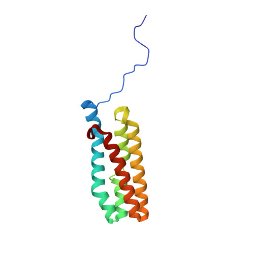
    In Synechocystis sp. PCC 6803, PsbQ is associated with photosystem II (PSII) complexes with the highest activity and stability. However, this subunit is not found in PSII X-ray crystallographic structures from Thermosynechococcus elongatus or Thermosynechococcus vulcanus. We present the crystal structure of cyanobacterial PsbQ determined in the presence and absence of Zn(2+). The protein has a well-defined helical core, containing four helices arranged in an up-down-up-down fold. A conserved potential interaction site composed of a divalent metal binding site and adjacent hydrophobic pocket has been identified.


  • Organizational Affiliation

    Department of Biochemistry, University of Otago, Dunedin 9054, New Zealand.


Macromolecules
Find similar proteins by:  (by identity cutoff)  |  3D Structure
Entity ID: 1
MoleculeChains Sequence LengthOrganismDetailsImage
Sll1638 protein
A, B
133Synechocystis sp. PCC 6803Mutation(s): 0 
Gene Names: sll1638
UniProt
Find proteins for P73048 (Synechocystis sp. (strain PCC 6803 / Kazusa))
Explore P73048 
Go to UniProtKB:  P73048
Entity Groups  
Sequence Clusters30% Identity50% Identity70% Identity90% Identity95% Identity100% Identity
UniProt GroupP73048
Sequence Annotations
Expand
  • Reference Sequence
Experimental Data & Validation

Experimental Data

  • Method: X-RAY DIFFRACTION
  • Resolution: 1.85 Å
  • R-Value Free: 0.218 
  • R-Value Work: 0.179 
  • R-Value Observed: 0.181 
  • Space Group: I 1 2 1
Unit Cell:
Length ( Å )Angle ( ˚ )
a = 92.771α = 90
b = 29.03β = 97.77
c = 99.503γ = 90
Software Package:
Software NamePurpose
SCALAdata scaling
PHASERphasing
REFMACrefinement
PDB_EXTRACTdata extraction
MOSFLMdata reduction

Structure Validation

View Full Validation Report


Entry History 

Deposition Data

Revision History  (Full details and data files)

  • Version 1.0: 2010-03-31
    Type: Initial release
  • Version 1.1: 2011-07-13
    Changes: Version format compliance
  • Version 1.2: 2023-11-01
    Changes: Data collection, Database references, Derived calculations, Refinement description