Limitations of peptide retro-inverso isomerization in molecular mimicry.
Li, C., Pazgier, M., Li, J., Li, C., Liu, M., Zou, G., Li, Z., Chen, J., Tarasov, S.G., Lu, W.Y., Lu, W.(2010) J Biol Chem 285: 19572-19581
- PubMed: 20382735
- DOI: https://doi.org/10.1074/jbc.M110.116814
- Primary Citation of Related Structures:
3LRY - PubMed Abstract:
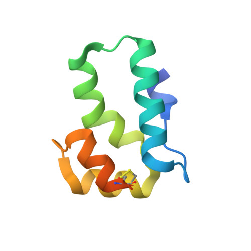
A retro-inverso peptide is made up of d-amino acids in a reversed sequence and, when extended, assumes a side chain topology similar to that of its parent molecule but with inverted amide peptide bonds. Despite their limited success as antigenic mimicry, retro-inverso isomers generally fail to emulate the protein-binding activities of their parent peptides of an alpha-helical nature. In studying the interaction between the tumor suppressor protein p53 and its negative regulator MDM2, Sakurai et al. (Sakurai, K., Chung, H. S., and Kahne, D. (2004) J. Am. Chem. Soc. 126, 16288-16289) made a surprising finding that the retro-inverso isomer of p53(15-29) retained the same binding activity as the wild type peptide as determined by inhibition enzyme-linked immunosorbent assay. The authors attributed the unusual outcome to the ability of the D-peptide to adopt a right-handed helical conformation upon MDM2 binding. Using a battery of biochemical and biophysical tools, we found that retro-inverso isomerization diminished p53 (15-29) binding to MDM2 or MDMX by 3.2-3.3 kcal/mol. Similar results were replicated with the C-terminal domain of HIV-1 capsid protein (3.0 kcal/mol) and the Src homology 3 domain of Abl tyrosine kinase (3.4 kcal/mol). CD and NMR spectroscopic as well as x-ray crystallographic studies showed that D-peptide ligands of MDM2 invariably adopted left-handed helical conformations in both free and bound states. Our findings reinforce that the retro-inverso strategy works poorly in molecular mimicry of biologically active helical peptides, due to inherent differences at the secondary and tertiary structure levels between an l-peptide and its retro-inverso isomer despite their similar side chain topologies at the primary structure level.
Organizational Affiliation:
School of Pharmacy, Fudan University, Shanghai 201203, China.