3LCB

The crystal structure of isocitrate dehydrogenase kinase/phosphatase in complex with its substrate, isocitrate dehydrogenase, from Escherichia coli.


Experimental Data Snapshot

  • Method: X-RAY DIFFRACTION
  • Resolution: 2.90 Å
  • R-Value Free: 0.222 
  • R-Value Work: 0.190 
  • R-Value Observed: 0.192 

wwPDB Validation   3D Report Full Report


Ligand Structure Quality Assessment 


This is version 1.3 of the entry. See complete history


Literature

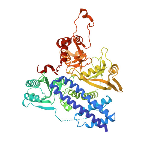
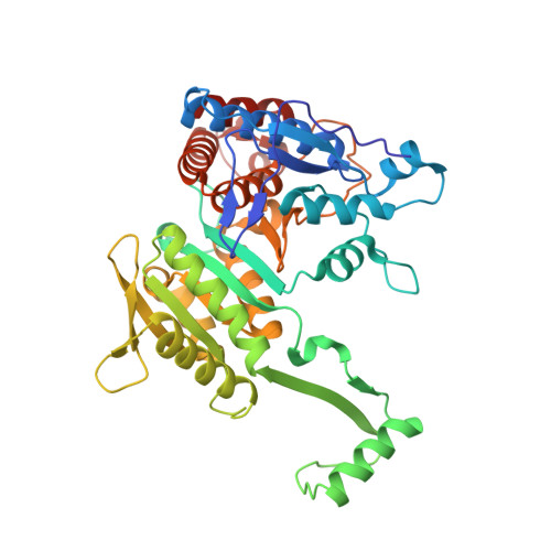
Structure of the bifunctional isocitrate dehydrogenase kinase/phosphatase.

Zheng, J.Jia, Z.

(2010) Nature 465: 961-965

  • DOI: https://doi.org/10.1038/nature09088
  • Primary Citation of Related Structures:  
    3EPS, 3LC6, 3LCB

  • PubMed Abstract: 

    The Escherichia coli isocitrate dehydrogenase kinase/phosphatase (AceK) is a unique bifunctional enzyme that phosphorylates or dephosphorylates isocitrate dehydrogenase (ICDH) in response to environmental changes, resulting in the inactivation or, respectively, activation of ICDH. ICDH inactivation short-circuits the Krebs cycle by enabling the glyoxlate bypass. It was the discovery of AceK and ICDH that established the existence of protein phosphorylation regulation in prokaryotes. As a 65-kDa protein, AceK is significantly larger than typical eukaryotic protein kinases. Apart from the ATP-binding motif, AceK does not share sequence homology with any eukaryotic protein kinase or phosphatase. Most intriguingly, AceK possesses the two opposing activities of protein kinase and phosphatase within one protein, and specifically recognizes only intact ICDH. Additionally, AceK has strong ATPase activity. It has been shown that AceK kinase, phosphatase and ATPase activities reside at the same site, although the molecular basis of such multifunctionality and its regulation remains completely unknown. Here we report the structures of AceK and its complex with ICDH. The AceK structure reveals a eukaryotic protein-kinase-like domain containing ATP and a regulatory domain with a novel fold. As an AceK phosphatase activator and kinase inhibitor, AMP is found to bind in an allosteric site between the two AceK domains. An AMP-mediated conformational change exposes and shields ATP, acting as a switch between AceK kinase and phosphatase activities, and ICDH-binding induces further conformational change for AceK activation. The substrate recognition loop of AceK binds to the ICDH dimer, allowing higher-order substrate recognition and interaction, and inducing critical conformational change at the phosphorylation site of ICDH.


  • Organizational Affiliation

    Department of Biochemistry, Queen's University, Kingston, Ontario K7L 3N6, Canada.


Macromolecules
Find similar proteins by:  (by identity cutoff)  |  3D Structure
Entity ID: 1
MoleculeChains Sequence LengthOrganismDetailsImage
Isocitrate dehydrogenase kinase/phosphatase
A, B
578Escherichia coli O157:H7Mutation(s): 0 
Gene Names: aceKECH74115_5487
EC: 2.7.11.5 (PDB Primary Data), 3.1.3 (PDB Primary Data)
UniProt
Find proteins for Q8X607 (Escherichia coli O157:H7)
Explore Q8X607 
Go to UniProtKB:  Q8X607
Entity Groups  
Sequence Clusters30% Identity50% Identity70% Identity90% Identity95% Identity100% Identity
UniProt GroupQ8X607
Sequence Annotations
Expand
  • Reference Sequence
Find similar proteins by:  (by identity cutoff)  |  3D Structure
Entity ID: 2
MoleculeChains Sequence LengthOrganismDetailsImage
Isocitrate dehydrogenase [NADP]
C, D
416Escherichia coli K-12Mutation(s): 0 
Gene Names: b1136icdicdAicdEicdhJW1122
EC: 1.1.1.42
UniProt
Find proteins for P08200 (Escherichia coli (strain K12))
Explore P08200 
Go to UniProtKB:  P08200
Entity Groups  
Sequence Clusters30% Identity50% Identity70% Identity90% Identity95% Identity100% Identity
UniProt GroupP08200
Sequence Annotations
Expand
  • Reference Sequence
Experimental Data & Validation

Experimental Data

  • Method: X-RAY DIFFRACTION
  • Resolution: 2.90 Å
  • R-Value Free: 0.222 
  • R-Value Work: 0.190 
  • R-Value Observed: 0.192 
  • Space Group: P 63
Unit Cell:
Length ( Å )Angle ( ˚ )
a = 196.801α = 90
b = 196.801β = 90
c = 156.458γ = 120
Software Package:
Software NamePurpose
DENZOdata reduction
SCALEPACKdata scaling
PHASERphasing
REFMACrefinement
PDB_EXTRACTdata extraction
HKL-2000data reduction
HKL-2000data scaling

Structure Validation

View Full Validation Report


Ligand Structure Quality Assessment 


Entry History 

Deposition Data

Revision History  (Full details and data files)

  • Version 1.0: 2010-04-21
    Type: Initial release
  • Version 1.1: 2011-07-13
    Changes: Version format compliance
  • Version 1.2: 2017-11-01
    Changes: Refinement description
  • Version 1.3: 2024-02-21
    Changes: Data collection, Database references, Derived calculations