3LAA

Crystal structure of the trimeric autotransporter adhesin head domain BpaA from Burkholderia pseudomallei


Experimental Data Snapshot

  • Method: X-RAY DIFFRACTION
  • Resolution: 1.35 Å
  • R-Value Free: 0.128 
  • R-Value Work: 0.114 
  • R-Value Observed: 0.115 

wwPDB Validation   3D Report Full Report


This is version 1.4 of the entry. See complete history


Literature

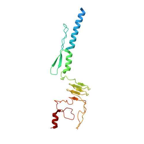
Structure of a Burkholderia pseudomallei trimeric autotransporter adhesin head.

Edwards, T.E.Phan, I.Abendroth, J.Dieterich, S.H.Masoudi, A.Guo, W.Hewitt, S.N.Kelley, A.Leibly, D.Brittnacher, M.J.Staker, B.L.Miller, S.I.Van Voorhis, W.C.Myler, P.J.Stewart, L.J.

(2010) PLoS One 5: 12803-12811

  • DOI: https://doi.org/10.1371/journal.pone.0012803
  • Primary Citation of Related Structures:  
    3LA9, 3LAA

  • PubMed Abstract: 

    Pathogenic bacteria adhere to the host cell surface using a family of outer membrane proteins called Trimeric Autotransporter Adhesins (TAAs). Although TAAs are highly divergent in sequence and domain structure, they are all conceptually comprised of a C-terminal membrane anchoring domain and an N-terminal passenger domain. Passenger domains consist of a secretion sequence, a head region that facilitates binding to the host cell surface, and a stalk region.


  • Organizational Affiliation

    Seattle Structural Genomics Center for Infectious Disease, Seattle, Washington, USA. tedwards@embios.com


Macromolecules
Find similar proteins by:  (by identity cutoff)  |  3D Structure
Entity ID: 1
MoleculeChains Sequence LengthOrganismDetailsImage
Haemagglutinin family protein200Burkholderia pseudomallei 1710bMutation(s): 0 
Gene Names: BURPS1710b_A0459xadA
UniProt
Find proteins for Q3JLD6 (Burkholderia pseudomallei (strain 1710b))
Explore Q3JLD6 
Go to UniProtKB:  Q3JLD6
Entity Groups  
Sequence Clusters30% Identity50% Identity70% Identity90% Identity95% Identity100% Identity
UniProt GroupQ3JLD6
Sequence Annotations
Expand
  • Reference Sequence
Experimental Data & Validation

Experimental Data

  • Method: X-RAY DIFFRACTION
  • Resolution: 1.35 Å
  • R-Value Free: 0.128 
  • R-Value Work: 0.114 
  • R-Value Observed: 0.115 
  • Space Group: H 3
  • Diffraction Data: https://doi.org/10.18430/M33LAA
Unit Cell:
Length ( Å )Angle ( ˚ )
a = 50.718α = 90
b = 50.718β = 90
c = 136.496γ = 120
Software Package:
Software NamePurpose
DENZOdata reduction
SCALEPACKdata scaling
PHASERphasing
REFMACrefinement
PDB_EXTRACTdata extraction
HKL-2000data reduction
HKL-2000data scaling

Structure Validation

View Full Validation Report


Entry History 

Deposition Data

Revision History  (Full details and data files)

  • Version 1.0: 2010-05-12
    Type: Initial release
  • Version 1.1: 2011-07-13
    Changes: Version format compliance
  • Version 1.2: 2017-11-01
    Changes: Refinement description
  • Version 1.3: 2018-04-18
    Changes: Data collection
  • Version 1.4: 2023-09-06
    Changes: Data collection, Database references, Refinement description