3L7R

crystal structure of MetE from streptococcus mutans


Experimental Data Snapshot

  • Method: X-RAY DIFFRACTION
  • Resolution: 2.40 Å
  • R-Value Free: 0.262 
  • R-Value Work: 0.200 
  • R-Value Observed: 0.203 

wwPDB Validation   3D Report Full Report


This is version 1.3 of the entry. See complete history


Literature

Crystal Structures of Cobalamin-Independent Methionine Synthase (MetE) from Streptococcus mutans: A Dynamic Zinc-Inversion Model

Fu, T.M.Almqvist, J.Liang, Y.H.Li, L.Huang, Y.Su, X.D.

(2011) J Mol Biol 412: 688-697

  • DOI: https://doi.org/10.1016/j.jmb.2011.08.005
  • Primary Citation of Related Structures:  
    3L7R, 3T0C

  • PubMed Abstract: 

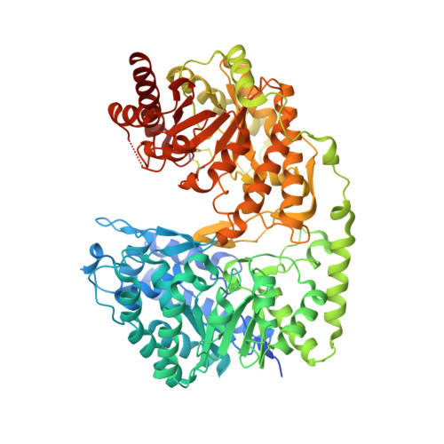
    Cobalamin-independent methionine synthase (MetE) catalyzes the direct transfer of a methyl group from methyltetrahydrofolate to l-homocysteine to form methionine. Previous studies have shown that the MetE active site coordinates a zinc atom, which is thought to act as a Lewis acid and plays a role in the activation of thiol. Extended X-ray absorption fine structure studies and mutagenesis experiments identified the zinc-binding site in MetE from Escherichia coli. Further structural investigations of MetE from Thermotoga maritima lead to the proposition of two models: "induced fit" and "dynamic equilibrium", to account for the catalytic mechanisms of MetE. Here, we present crystal structures of oxidized and zinc-replete MetE from Streptococcus mutans at the physiological pH. The structures reveal that zinc is mobile in the active center and has the possibility to invert even in the absence of homocysteine. These structures provide evidence for the dynamic equilibrium model.


  • Organizational Affiliation

    State Key Laboratory of Protein and Plant Gene Research, School of Life Sciences, Peking University, Beijing 100871, PR China.


Macromolecules
Find similar proteins by:  (by identity cutoff)  |  3D Structure
Entity ID: 1
MoleculeChains Sequence LengthOrganismDetailsImage
5-methyltetrahydropteroyltriglutamate--homocysteine methyltransferase779Streptococcus mutans UA159Mutation(s): 0 
Gene Names: SMU_873
EC: 2.1.1.14
UniProt
Find proteins for Q8CWX6 (Streptococcus mutans serotype c (strain ATCC 700610 / UA159))
Explore Q8CWX6 
Go to UniProtKB:  Q8CWX6
Entity Groups  
Sequence Clusters30% Identity50% Identity70% Identity90% Identity95% Identity100% Identity
UniProt GroupQ8CWX6
Sequence Annotations
Expand
  • Reference Sequence
Experimental Data & Validation

Experimental Data

  • Method: X-RAY DIFFRACTION
  • Resolution: 2.40 Å
  • R-Value Free: 0.262 
  • R-Value Work: 0.200 
  • R-Value Observed: 0.203 
  • Space Group: P 1 21 1
Unit Cell:
Length ( Å )Angle ( ˚ )
a = 52.913α = 90
b = 99.36β = 94.95
c = 77.448γ = 90
Software Package:
Software NamePurpose
PROTEUM PLUSdata collection
MOLREPphasing
PHENIXrefinement
PROTEUM PLUSdata reduction
PROTEUM PLUSdata scaling

Structure Validation

View Full Validation Report


Entry History 

Deposition Data

Revision History  (Full details and data files)

  • Version 1.0: 2011-01-12
    Type: Initial release
  • Version 1.1: 2011-07-13
    Changes: Version format compliance
  • Version 1.2: 2011-09-28
    Changes: Database references
  • Version 1.3: 2023-11-01
    Changes: Data collection, Database references, Derived calculations, Refinement description