1-(3-Deoxy-3-fluoro-beta-d-glucopyranosyl) pyrimidine derivatives as inhibitors of glycogen phosphorylase b: Kinetic, crystallographic and modelling studies.
Tsirkone, V.G., Tsoukala, E., Lamprakis, C., Manta, S., Hayes, J.M., Skamnaki, V.T., Drakou, C., Zographos, S.E., Komiotis, D., Leonidas, D.D.(2010) Bioorg Med Chem 18: 3413-3425
- PubMed: 20430629
- DOI: https://doi.org/10.1016/j.bmc.2010.04.004
- Primary Citation of Related Structures:
3L79, 3L7A, 3L7B, 3L7C, 3L7D - PubMed Abstract:
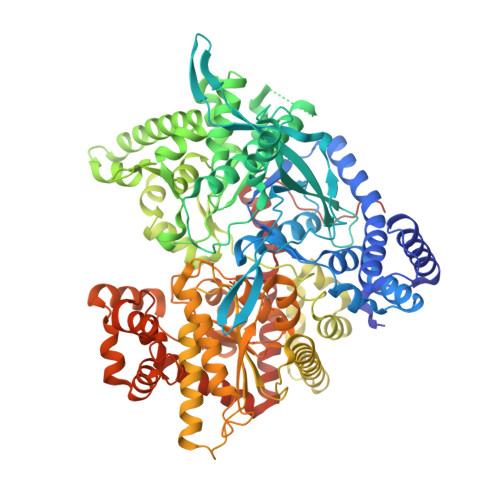
Design of inhibitors of glycogen phosphorylase (GP) with pharmaceutical applications in improving glycaemic control in type 2 diabetes is a promising therapeutic strategy. The catalytic site of muscle glycogen phosphorylase b (GPb) has been probed with five deoxy-fluro-glucose derivatives. These inhibitors had fluorine instead of hydroxyl at the 3' position of the glucose moiety and a variety of pyrimidine derivatives at the 1' position. The best of this carbohydrate-based family of five inhibitors displays a K(i) value of 46muM. To elucidate the mechanism of inhibition for these compounds, the crystal structures of GPb in complex with each ligand were determined and refined to high resolution. The structures demonstrated that the inhibitors bind preferentially at the catalytic site and promote the less active T state conformation of the enzyme by making several favorable contacts with residues of the 280s loop. Fluorine is engaged in hydrogen bond interactions but does not improve glucose potency. The pyrimidine groups are located between residues 284-286 of the 280s loop, Ala383 of the 380s loop, and His341 of the beta-pocket. These interactions appear important in stabilizing the inactive quaternary T state of the enzyme. As a follow up to recent computations performed on beta-d-glucose pyrimidine derivatives, tautomeric forms of ligands 1-5 were considered as potential binding states. Using Glide-XP docking and QM/MM calculations, the ligands 2 and 5 are predicted to bind in different tautomeric states in their respective GPb complexes. Also, using alpha-d-glucose as a benchmark model, a series of substitutions for glucose -OH at the 3' (equatorial) position were investigated for their potential to improve the binding affinity of glucose-based GPb catalytic site inhibitors. Glide-XP and quantum mechanics polarized ligand (QPLD-SP/XP) docking calculations revealed favorable binding at this position to be dominated by hydrogen bond contributions; none of the substitutions (including fluorine) out-performed the native -OH substituent which can act both as hydrogen bond donor and acceptor. The structural analyses of these compounds can be exploited towards the development of better inhibitors.
Organizational Affiliation:
Institute of Organic & Pharmaceutical Chemistry, National Hellenic Research Foundation, 48 Vas. Constantinou Avenue, 11635 Athens, Greece.