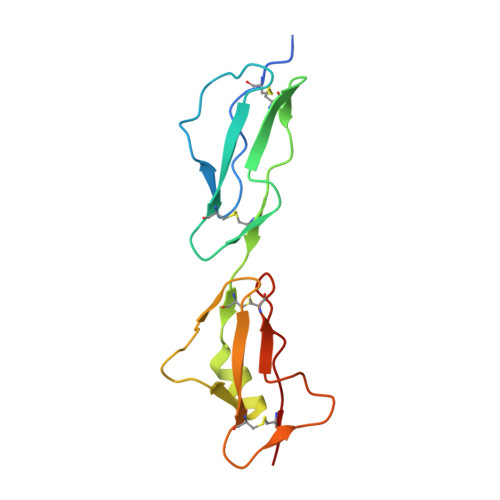
Both domain 19 and domain 20 of factor H are involved in binding to complement C3b and C3d
Bhattacharjee, A., Lehtinen, M.J., Kajander, T., Goldman, A., Jokiranta, T.S.(2010) Mol Immunol 47: 1686-1691
- PubMed: 20378178
- DOI: https://doi.org/10.1016/j.molimm.2010.03.007
- Primary Citation of Related Structures:
3KXV, 3KZJ - PubMed Abstract:
Factor H (FH) regulates the alternative pathway of complement in plasma and mediates discrimination of cellular surfaces to alternative pathway activators and non-activators. The carboxyl-terminal domains 19 and 20 of FH are essential in target discrimination and are known to contain binding sites for the C3d part of C3b, heparin, and endothelial cells. Mutations in FH19-20 are frequently found in patients with atypical haemolytic uremic syndrome (aHUS). Most aHUS-associated and some other mutations have been shown to lead to impaired binding to C3d and C3b by the recombinant FH19-20 fragment. Most of these mutated residues, such as R1203, are located close to each other in domain 20 but some, such as Q1139, are located in domain 19. We generated mutant proteins Q1139A and R1203A of FH19-20 and showed that their binding to C3d and C3b was clearly impaired. To show that the effects on C3d/C3b binding are due to direct interactions rather than structural changes, we solved the X-ray crystal structures of the R1203A and Q1139A mutant proteins at 1.65 and 2.0A, respectively. Neither of the mutations caused any overall structural changes in FH19-20. It is thus evident that Q1139 in domain 19 and R1203 in domain 20 are directly involved in binding to the C3d part of C3b and therefore both the domains are involved in the interaction with C3d and C3b. This explains why several aHUS-associated FH mutations are found within domain 19 in addition to domain 20.
Organizational Affiliation:
Department of Bacteriology and Immunology, Haartman Institute, University of Helsinki, Haartmaninkatu 3, FIN-00014, Finland.