3KP1

Crystal structure of ornithine 4,5 aminomutase (Resting State)


Experimental Data Snapshot

  • Method: X-RAY DIFFRACTION
  • Resolution: 2.01 Å
  • R-Value Free: 0.216 
  • R-Value Work: 0.163 
  • R-Value Observed: 0.166 

wwPDB Validation   3D Report Full Report


Ligand Structure Quality Assessment 


This is version 1.4 of the entry. See complete history


Literature

Large-scale domain dynamics and adenosylcobalamin reorientation orchestrate radical catalysis in ornithine 4,5-aminomutase.

Wolthers, K.R.Levy, C.Scrutton, N.S.Leys, D.

(2010) J Biol Chem 285: 13942-13950

  • DOI: https://doi.org/10.1074/jbc.M109.068908
  • Primary Citation of Related Structures:  
    3KOW, 3KOX, 3KOY, 3KOZ, 3KP0, 3KP1

  • PubMed Abstract: 

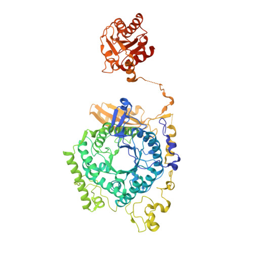
    D-ornithine 4,5-aminomutase (OAM) from Clostridium sticklandii converts D-ornithine to 2,4-diaminopentanoic acid by way of radical propagation from an adenosylcobalamin (AdoCbl) to a pyridoxal 5'-phosphate (PLP) cofactor. We have solved OAM crystal structures in different catalytic states that together demonstrate unusual stability of the AdoCbl Co-C bond and that radical catalysis is coupled to large-scale domain motion. The 2.0-A substrate-free enzyme crystal structure reveals the Rossmann domain, harboring the intact AdoCbl cofactor, is tilted toward the edge of the PLP binding triose-phosphate isomerase barrel domain. The PLP forms an internal aldimine link to the Rossmann domain through Lys(629), effectively locking the enzyme in this "open" pre-catalytic conformation. The distance between PLP and 5'-deoxyadenosyl group is 23 A, and large-scale domain movement is thus required prior to radical catalysis. The OAM crystals contain two Rossmann domains within the asymmetric unit that are unconstrained by the crystal lattice. Surprisingly, the binding of various ligands to OAM crystals (in an oxygen-free environment) leads to transimination in the absence of significant reorientation of the Rossmann domains. In contrast, when performed under aerobic conditions, this leads to extreme disorder in the latter domains correlated with the loss of the 5'-deoxyadenosyl group. Our data indicate turnover and hence formation of the "closed" conformation is occurring within OAM crystals, but that the equilibrium is poised toward the open conformation. We propose that substrate binding induces large-scale domain motion concomitant with a reconfiguration of the 5'-deoxyadenosyl group, triggering radical catalysis in OAM.


  • Organizational Affiliation

    Faculty of Life Sciences, University of Manchester, Manchester Interdisciplinary Biocentre, 131 Princess Street, Manchester M1 7DN, United Kingdom. kirsten.wolthers@manchester.ac.uk


Macromolecules
Find similar proteins by:  (by identity cutoff)  |  3D Structure
Entity ID: 1
MoleculeChains Sequence LengthOrganismDetailsImage
D-ornithine aminomutase E componentA,
C [auth B],
E [auth D],
G [auth C]
763Acetoanaerobium sticklandiiMutation(s): 0 
Gene Names: oraE
UniProt
Find proteins for E3PY95 (Acetoanaerobium sticklandii (strain ATCC 12662 / DSM 519 / JCM 1433 / CCUG 9281 / NCIMB 10654 / HF))
Explore E3PY95 
Go to UniProtKB:  E3PY95
Entity Groups  
Sequence Clusters30% Identity50% Identity70% Identity90% Identity95% Identity100% Identity
UniProt GroupE3PY95
Sequence Annotations
Expand
  • Reference Sequence
Find similar proteins by:  (by identity cutoff)  |  3D Structure
Entity ID: 2
MoleculeChains Sequence LengthOrganismDetailsImage
D-ornithine aminomutase S componentB [auth E],
D [auth F],
F [auth H],
H [auth G]
121Acetoanaerobium sticklandiiMutation(s): 0 
Gene Names: oraS
UniProt
Find proteins for E3PY96 (Acetoanaerobium sticklandii (strain ATCC 12662 / DSM 519 / JCM 1433 / CCUG 9281 / NCIMB 10654 / HF))
Explore E3PY96 
Go to UniProtKB:  E3PY96
Entity Groups  
Sequence Clusters30% Identity50% Identity70% Identity90% Identity95% Identity100% Identity
UniProt GroupE3PY96
Sequence Annotations
Expand
  • Reference Sequence
Small Molecules
Ligands 3 Unique
IDChains Name / Formula / InChI Key2D Diagram3D Interactions
B12
Query on B12

Download Ideal Coordinates CCD File 
J [auth A],
M [auth B],
P [auth D],
S [auth C]
COBALAMIN
C62 H89 Co N13 O14 P
LKVIQTCSMMVGFU-DWSMJLPVSA-N
5AD
Query on 5AD

Download Ideal Coordinates CCD File 
K [auth A],
N [auth B],
Q [auth D],
T [auth C]
5'-DEOXYADENOSINE
C10 H13 N5 O3
XGYIMTFOTBMPFP-KQYNXXCUSA-N
PLP
Query on PLP

Download Ideal Coordinates CCD File 
I [auth A],
L [auth B],
O [auth D],
R [auth C]
PYRIDOXAL-5'-PHOSPHATE
C8 H10 N O6 P
NGVDGCNFYWLIFO-UHFFFAOYSA-N
Experimental Data & Validation

Experimental Data

  • Method: X-RAY DIFFRACTION
  • Resolution: 2.01 Å
  • R-Value Free: 0.216 
  • R-Value Work: 0.163 
  • R-Value Observed: 0.166 
  • Space Group: P 1 21 1
Unit Cell:
Length ( Å )Angle ( ˚ )
a = 66.43α = 90
b = 234.51β = 103.51
c = 124.56γ = 90
Software Package:
Software NamePurpose
PHENIXrefinement
PHASERphasing
REFMACrefinement
MOSFLMdata reduction
SCALAdata scaling
DNAdata collection

Structure Validation

View Full Validation Report


Ligand Structure Quality Assessment 


Entry History 

Deposition Data

Revision History  (Full details and data files)

  • Version 1.0: 2010-01-26
    Type: Initial release
  • Version 1.1: 2011-07-13
    Changes: Version format compliance
  • Version 1.2: 2012-10-24
    Changes: Non-polymer description
  • Version 1.3: 2019-07-17
    Changes: Data collection, Derived calculations, Refinement description
  • Version 1.4: 2019-10-30
    Changes: Advisory, Data collection, Database references, Derived calculations