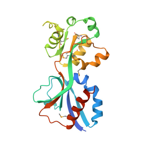
Crystal Structure of the AmpR Effector Binding Domain Provides Insight into the Molecular Regulation of Inducible AmpC beta-Lactamase.
Balcewich, M.D., Reeve, T.M., Orlikow, E.A., Donald, L.J., Vocadlo, D.J., Mark, B.L.(2010) J Mol Biol 400: 998-1010
- PubMed: 20594961
- DOI: https://doi.org/10.1016/j.jmb.2010.05.040
- Primary Citation of Related Structures:
3KOS, 3KOT - PubMed Abstract:
Hyperproduction of AmpC beta-lactamase (AmpC) is a formidable mechanism of resistance to penicillins and cephalosporins in Gram-negative bacteria such as Pseudomonas aeruginosa and Enterobacteriaceae. AmpC expression is regulated by the LysR-type transcriptional regulator AmpR. ampR and ampC genes form a divergent operon with overlapping promoters to which AmpR binds and regulates the transcription of both genes. AmpR induces ampC by binding to one member of the family of 1,6-anhydro-N-acetylmuramyl peptides, which are cytosolic catabolites of peptidoglycan that accumulate during beta-lactam challenge. To gain structural insights into AmpR regulation, we determined the crystal structure of the effector binding domain (EBD) of AmpR from Citrobacter freundii up to 1.83 A resolution. The AmpR EBD is dimeric and each monomer comprises two subdomains that adopt alpha/beta Rossmann-like folds. Located between the monomer subdomains is a pocket that was found to bind the crystallization buffer molecule 2-(N-morpholino)ethanesulfonic acid. The pocket, together with a groove along the surface of subdomain I, forms a putative effector binding site into which a molecule of 1,6-anhydro-N-acetylmuramyl pentapeptide could be modeled. Amino acid substitutions at the base of the interdomain pocket either were found to render AmpR incapable of inducing ampC (Thr103Val, Ser221Ala and Tyr264Phe) or resulted in constitutive ampC expression (Gly102Glu). While the substitutions that prevented ampC induction did not alter the overall AmpR EBD structure, circular dichroism spectroscopy revealed that the nonconservative Gly102Glu mutation affected EBD secondary structure, confirming previous work suggesting that Gly102Glu induces a conformational change to result in constitutive AmpC production.
Organizational Affiliation:
Department of Microbiology, University of Manitoba, 418 Buller Building, Winnipeg, Manitoba, Canada R3T 2N2.