Mutation of archaeal isopentenyl phosphate kinase highlights mechanism and guides phosphorylation of additional isoprenoid monophosphates.
Dellas, N., Noel, J.P.(2010) ACS Chem Biol 5: 589-601
- PubMed: 20392112
- DOI: https://doi.org/10.1021/cb1000313
- Primary Citation of Related Structures:
3K4O, 3K4Y, 3K52, 3K56 - PubMed Abstract:
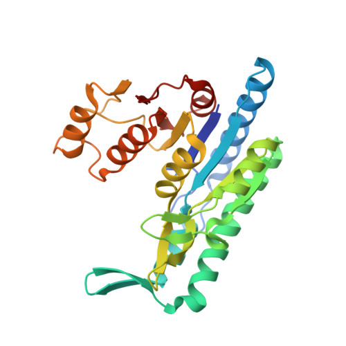
The biosynthesis of isopentenyl diphosphate (IPP) from either the mevalonate (MVA) or the 1-deoxy-d-xylulose 5-phosphate (DXP) pathway provides the key metabolite for primary and secondary isoprenoid biosynthesis. Isoprenoid metabolism plays crucial roles in membrane stability, steroid biosynthesis, vitamin production, protein localization, defense and communication, photoprotection, sugar transport, and glycoprotein biosynthesis. Recently, an alternative branch of the MVA pathway was discovered in the archaeon Methanocaldococcus jannaschii involving a small molecule kinase, isopentenyl phosphate kinase (IPK). IPK belongs to the amino acid kinase (AAK) superfamily. In vitro, IPK phosphorylates isopentenyl monophosphate (IP) in an ATP and Mg(2+)-dependent reaction producing IPP. Here, we describe crystal structures of IPK from M. jannaschii refined to nominal resolutions of 2.0-2.8 A. Notably, an active site histidine residue (His60) forms a hydrogen bond with the terminal phosphate of both substrate and product. This His residue serves as a marker for a subset of the AAK family that catalyzes phosphorylation of phosphate or phosphonate functional groups; the larger family includes carboxyl-directed kinases, which lack this active site residue. Using steady-state kinetic analysis of H60A, H60N, and H60Q mutants, the protonated form of the Nepsilon(2) nitrogen of His60 was shown to be essential for catalysis, most likely through hydrogen bond stabilization of the transition state accompanying transphosphorylation. Moreover, the structures served as the starting point for the engineering of IPK mutants capable of the chemoenzymatic synthesis of longer chain isoprenoid diphosphates from monophosphate precursors.
Organizational Affiliation:
Salk Institute for Biological Studies, Jack H. Skirball Center for Chemical Biology and Proteomics, La Jolla, California 92037, USA.