3JQM

Binding of 5'-GTP to molybdenum cofactor biosynthesis protein MoaC from Thermus theromophilus HB8


Experimental Data Snapshot

  • Method: X-RAY DIFFRACTION
  • Resolution: 2.50 Å
  • R-Value Free: 0.270 
  • R-Value Work: 0.202 
  • R-Value Observed: 0.202 

wwPDB Validation   3D Report Full Report


Ligand Structure Quality Assessment 


This is version 1.3 of the entry. See complete history


Literature

Structures of apo and GTP-bound molybdenum cofactor biosynthesis protein MoaC from Thermus thermophilus HB8

Kanaujia, S.P.Jeyakanthan, J.Nakagawa, N.Balasubramaniam, S.Shinkai, A.Kuramitsu, S.Yokoyama, S.Sekar, K.

(2010) Acta Crystallogr D Biol Crystallogr 66: 821-833

  • DOI: https://doi.org/10.1107/S0907444910019074
  • Primary Citation of Related Structures:  
    3JQJ, 3JQK, 3JQM

  • PubMed Abstract: 

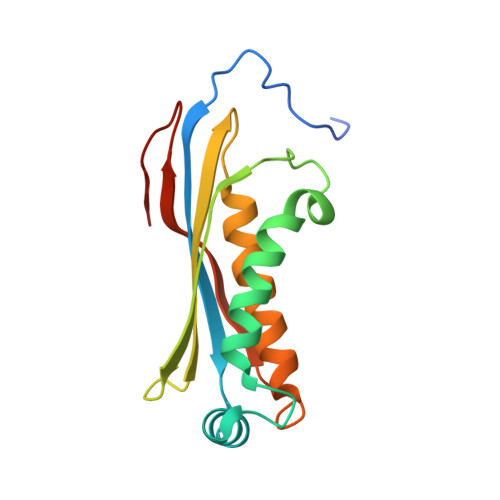
    The first step in the molybdenum cofactor (Moco) biosynthesis pathway involves the conversion of guanosine triphosphate (GTP) to precursor Z by two proteins (MoaA and MoaC). MoaA belongs to the S-adenosylmethionine-dependent radical enzyme superfamily and is believed to generate protein and/or substrate radicals by reductive cleavage of S-adenosylmethionine using an Fe-S cluster. MoaC has been suggested to catalyze the release of pyrophosphate and the formation of the cyclic phosphate of precursor Z. However, structural evidence showing the binding of a substrate-like molecule to MoaC is not available. Here, apo and GTP-bound crystal structures of MoaC from Thermus thermophilus HB8 are reported. Furthermore, isothermal titration calorimetry experiments have been carried out in order to obtain thermodynamic parameters for the protein-ligand interactions. In addition, molecular-dynamics (MD) simulations have been carried out on the protein-ligand complex of known structure and on models of relevant complexes for which X-ray structures are not available. The biophysical, structural and MD results reveal the residues that are involved in substrate binding and help in speculating upon a possible mechanism.


  • Organizational Affiliation

    Bioinformatics Centre (Centre of Excellence in Structural Biology and Bio-computing), Indian Institute of Science, Bangalore, India.


Macromolecules
Find similar proteins by:  (by identity cutoff)  |  3D Structure
Entity ID: 1
MoleculeChains Sequence LengthOrganismDetailsImage
Molybdenum cofactor biosynthesis protein C
A, B, C, D, E
A, B, C, D, E, F, G, H, I
157Thermus thermophilus HB8Mutation(s): 0 
Gene Names: TTHA1789
UniProt
Find proteins for Q5SHE1 (Thermus thermophilus (strain ATCC 27634 / DSM 579 / HB8))
Explore Q5SHE1 
Go to UniProtKB:  Q5SHE1
Entity Groups  
Sequence Clusters30% Identity50% Identity70% Identity90% Identity95% Identity100% Identity
UniProt GroupQ5SHE1
Sequence Annotations
Expand
  • Reference Sequence
Small Molecules
Ligands 5 Unique
IDChains Name / Formula / InChI Key2D Diagram3D Interactions
GTP
Query on GTP

Download Ideal Coordinates CCD File 
BB [auth I]
CA [auth D]
GA [auth E]
J [auth A]
JA [auth F]
BB [auth I],
CA [auth D],
GA [auth E],
J [auth A],
JA [auth F],
PA [auth G],
R [auth B],
VA [auth H],
X [auth C]
GUANOSINE-5'-TRIPHOSPHATE
C10 H16 N5 O14 P3
XKMLYUALXHKNFT-UUOKFMHZSA-N
FLC
Query on FLC

Download Ideal Coordinates CCD File 
CB [auth I]
DA [auth D]
HA [auth E]
K [auth A]
KA [auth F]
CB [auth I],
DA [auth D],
HA [auth E],
K [auth A],
KA [auth F],
QA [auth G],
S [auth B],
WA [auth H],
Y [auth C]
CITRATE ANION
C6 H5 O7
KRKNYBCHXYNGOX-UHFFFAOYSA-K
PEG
Query on PEG

Download Ideal Coordinates CCD File 
AB [auth H]
BA [auth C]
IA [auth E]
OA [auth F]
P [auth A]
AB [auth H],
BA [auth C],
IA [auth E],
OA [auth F],
P [auth A],
Q [auth A],
W [auth B]
DI(HYDROXYETHYL)ETHER
C4 H10 O3
MTHSVFCYNBDYFN-UHFFFAOYSA-N
GOL
Query on GOL

Download Ideal Coordinates CCD File 
L [auth A],
LA [auth F],
M [auth A],
XA [auth H],
Z [auth C]
GLYCEROL
C3 H8 O3
PEDCQBHIVMGVHV-UHFFFAOYSA-N
EDO
Query on EDO

Download Ideal Coordinates CCD File 
AA [auth C]
DB [auth I]
EA [auth D]
EB [auth I]
FA [auth D]
AA [auth C],
DB [auth I],
EA [auth D],
EB [auth I],
FA [auth D],
MA [auth F],
N [auth A],
NA [auth F],
O [auth A],
RA [auth G],
SA [auth G],
T [auth B],
TA [auth G],
U [auth B],
UA [auth G],
V [auth B],
YA [auth H],
ZA [auth H]
1,2-ETHANEDIOL
C2 H6 O2
LYCAIKOWRPUZTN-UHFFFAOYSA-N
Binding Affinity Annotations 
IDSourceBinding Affinity
GTP Binding MOAD:  3JQM Kd: 4.44e+4 (nM) from 1 assay(s)
Experimental Data & Validation

Experimental Data

  • Method: X-RAY DIFFRACTION
  • Resolution: 2.50 Å
  • R-Value Free: 0.270 
  • R-Value Work: 0.202 
  • R-Value Observed: 0.202 
  • Space Group: C 2 2 21
Unit Cell:
Length ( Å )Angle ( ˚ )
a = 69.929α = 90
b = 111.567β = 90
c = 311.423γ = 90
Software Package:
Software NamePurpose
HKL-2000data collection
PHASERphasing
CNSrefinement
HKL-2000data reduction
HKL-2000data scaling

Structure Validation

View Full Validation Report


Ligand Structure Quality Assessment 


Entry History 

Revision History  (Full details and data files)

  • Version 1.0: 2010-06-30
    Type: Initial release
  • Version 1.1: 2011-07-13
    Changes: Version format compliance
  • Version 1.2: 2012-04-11
    Changes: Database references
  • Version 1.3: 2023-11-01
    Changes: Data collection, Database references, Derived calculations, Refinement description