3IT6

The Crystal Structure of Ornithine Acetyltransferase complexed with Ornithine from Mycobacterium tuberculosis (Rv1653) at 2.4 A


Experimental Data Snapshot

  • Method: X-RAY DIFFRACTION
  • Resolution: 2.40 Å
  • R-Value Free: 0.267 
  • R-Value Work: 0.210 
  • R-Value Observed: 0.212 

wwPDB Validation   3D Report Full Report


This is version 1.3 of the entry. See complete history


Literature

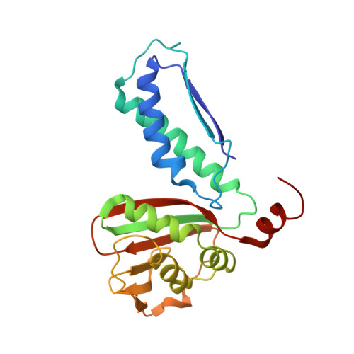
The molecular structure of ornithine acetyltransferase from Mycobacterium tuberculosis bound to ornithine, a competitive inhibitor.

Sankaranarayanan, R.Cherney, M.M.Garen, C.Garen, G.Niu, C.Yuan, M.James, M.N.

(2010) J Mol Biol 397: 979-990

  • DOI: https://doi.org/10.1016/j.jmb.2010.02.018
  • Primary Citation of Related Structures:  
    3IT4, 3IT6

  • PubMed Abstract: 

    Mycobacterium tuberculosis ornithine acetyltransferase (Mtb OAT; E.C. 2.3.1.35) is a key enzyme of the acetyl recycling pathway during arginine biosynthesis. It reversibly catalyzes the transfer of the acetyl group from N-acetylornithine (NAORN) to L-glutamate. Mtb OAT is a member of the N-terminal nucleophile fold family of enzymes. The crystal structures of Mtb OAT in native form and in its complex with ornithine (ORN) have been determined at 1.7 and 2.4 A resolutions, respectively. ORN is a competitive inhibitor of this enzyme against L-glutamate as substrate. Although the acyl-enzyme complex of Streptomyces clavuligerus ornithine acetyltransferase has been determined, ours is the first crystal structure to be reported of an ornithine acetyltransferase in complex with an inhibitor. ORN binding does not alter the structure of Mtb OAT globally. However, its presence stabilizes the three C-terminal residues that are disordered and not observed in the native structure. Also, stabilization of the C-terminal residues by ORN reduces the size of the active-site pocket volume in the structure of the ORN complex. The interactions of ORN and the protein residues of Mtb OAT unambiguously delineate the active-site residues of this enzyme in Mtb. Moreover, modeling studies carried out with NAORN based on the structure of the ORN-Mtb OAT complex reveal important interactions of the carbonyl oxygen of the acetyl group of NAORN with the main-chain nitrogen atom of Gly128 and with the side-chain oxygen of Thr127. These interactions likely help in the stabilization of oxyanion formation during enzymatic reaction and also will polarize the carbonyl carbon-oxygen bond, thereby enabling the side-chain atom O(gamma 1) of Thr200 to launch a nucleophilic attack on the carbonyl-carbon atom of the acetyl group of NAORN.


  • Organizational Affiliation

    Group in Protein Structure and Function, Department of Biochemistry, School of Molecular and Systems Medicine, Faculty of Medicine and Dentistry, University of Alberta, Edmonton, Alberta, Canada.


Macromolecules
Find similar proteins by:  (by identity cutoff)  |  3D Structure
Entity ID: 1
MoleculeChains Sequence LengthOrganismDetailsImage
Arginine biosynthesis bifunctional protein argJ alpha chain
A, C
199Mycobacterium tuberculosisMutation(s): 0 
Gene Names: argJMT1691MTCY06H11.18Rv1653
EC: 2.3.1.35
UniProt
Find proteins for P9WPZ3 (Mycobacterium tuberculosis (strain ATCC 25618 / H37Rv))
Explore P9WPZ3 
Go to UniProtKB:  P9WPZ3
Entity Groups  
Sequence Clusters30% Identity50% Identity70% Identity90% Identity95% Identity100% Identity
UniProt GroupP9WPZ3
Sequence Annotations
Expand
  • Reference Sequence
Find similar proteins by:  (by identity cutoff)  |  3D Structure
Entity ID: 2
MoleculeChains Sequence LengthOrganismDetailsImage
Arginine biosynthesis bifunctional protein argJ beta chain
B, D
205Mycobacterium tuberculosisMutation(s): 0 
Gene Names: argJRv1653MT1691MTCY06H11.18
EC: 2.3.1.1
UniProt
Find proteins for P9WPZ3 (Mycobacterium tuberculosis (strain ATCC 25618 / H37Rv))
Explore P9WPZ3 
Go to UniProtKB:  P9WPZ3
Entity Groups  
Sequence Clusters30% Identity50% Identity70% Identity90% Identity95% Identity100% Identity
UniProt GroupP9WPZ3
Sequence Annotations
Expand
  • Reference Sequence
Experimental Data & Validation

Experimental Data

  • Method: X-RAY DIFFRACTION
  • Resolution: 2.40 Å
  • R-Value Free: 0.267 
  • R-Value Work: 0.210 
  • R-Value Observed: 0.212 
  • Space Group: P 21 21 21
Unit Cell:
Length ( Å )Angle ( ˚ )
a = 60.849α = 90
b = 100.095β = 90
c = 156.401γ = 90
Software Package:
Software NamePurpose
ADSCdata collection
PHASERphasing
REFMACrefinement
HKL-2000data reduction
HKL-2000data scaling

Structure Validation

View Full Validation Report


Entry History 

Deposition Data

Revision History  (Full details and data files)

  • Version 1.0: 2010-03-02
    Type: Initial release
  • Version 1.1: 2011-07-13
    Changes: Version format compliance
  • Version 1.2: 2014-06-04
    Changes: Data collection
  • Version 1.3: 2023-09-06
    Changes: Data collection, Database references, Derived calculations, Refinement description