3I6B

Crystal structure of YrbI lacking the last 8 residues, in complex with Kdo and inorganic phosphate


Experimental Data Snapshot

  • Method: X-RAY DIFFRACTION
  • Resolution: 2.49 Å
  • R-Value Free: 0.238 
  • R-Value Work: 0.200 
  • R-Value Observed: 0.202 

wwPDB Validation   3D Report Full Report


Ligand Structure Quality Assessment 


This is version 1.3 of the entry. See complete history


Literature

The Tail of KdsC: CONFORMATIONAL CHANGES CONTROL THE ACTIVITY OF A HALOACID DEHALOGENASE SUPERFAMILY PHOSPHATASE.

Biswas, T.Yi, L.Aggarwal, P.Wu, J.Rubin, J.R.Stuckey, J.A.Woodard, R.W.Tsodikov, O.V.

(2009) J Biol Chem 284: 30594-30603

  • DOI: https://doi.org/10.1074/jbc.M109.012278
  • Primary Citation of Related Structures:  
    2R8E, 2R8X, 2R8Y, 2R8Z, 3HYC, 3I6B

  • PubMed Abstract: 

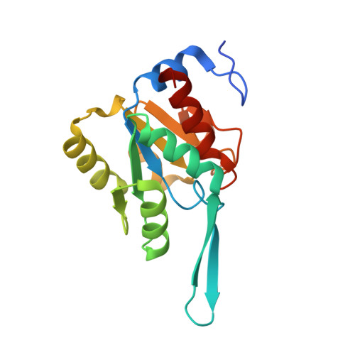
    The phosphatase KdsC cleaves 3-deoxy-D-manno-octulosonate 8-phosphate to generate a molecule of inorganic phosphate and Kdo. Kdo is an essential component of the lipopolysaccharide envelope in Gram-negative bacteria. Because lipopolysaccharide is an important determinant of bacterial resistance and toxicity, KdsC is a potential target for novel antibacterial agents. KdsC belongs to the broad haloacid dehalogenase superfamily. In haloacid dehalogenase superfamily enzymes, substrate specificity and catalytic efficiency are generally dictated by a fold feature called the cap domain. It is therefore not clear why KdsC, which lacks a cap domain, is catalytically efficient and highly specific to 3-deoxy-D-manno-octulosonate 8-phosphate. Here, we present a set of seven structures of tetrameric Escherichia coli KdsC (ranging from 1.4 to 3.06 A in resolution) that model different intermediate states in its catalytic mechanism. A crystal structure of product-bound E. coli KdsC shows how the interface between adjacent monomers defines the active site pocket. Kdo is engaged in a network of polar and nonpolar interactions with residues at this interface, which explains substrate specificity. Furthermore, this structural and kinetic analysis strongly suggests that the binding of the flexible C-terminal region (tail) to the active site makes KdsC catalytically efficient by facilitating product release.


  • Organizational Affiliation

    Department of Medicinal Chemistry, College of Pharmacy, University of Michigan, Ann Arbor, Michigan 48109, USA.


Macromolecules
Find similar proteins by:  (by identity cutoff)  |  3D Structure
Entity ID: 1
MoleculeChains Sequence LengthOrganismDetailsImage
3-deoxy-D-manno-octulosonate 8-phosphate phosphatase
A, B, C, D
180Escherichia coli BMutation(s): 0 
Gene Names: c3958kdsCYrbI
EC: 3.1.3.45
UniProt
Find proteins for P67653 (Escherichia coli O6:H1 (strain CFT073 / ATCC 700928 / UPEC))
Explore P67653 
Go to UniProtKB:  P67653
Entity Groups  
Sequence Clusters30% Identity50% Identity70% Identity90% Identity95% Identity100% Identity
UniProt GroupP67653
Sequence Annotations
Expand
  • Reference Sequence
Small Molecules
Ligands 3 Unique
IDChains Name / Formula / InChI Key2D Diagram3D Interactions
KDO
Query on KDO

Download Ideal Coordinates CCD File 
I [auth B],
N [auth D]
3-deoxy-alpha-D-manno-oct-2-ulopyranosonic acid
C8 H14 O8
NNLZBVFSCVTSLA-HXUQBWEZSA-N
PO4
Query on PO4

Download Ideal Coordinates CCD File 
F [auth A],
H [auth B],
K [auth C],
M [auth D]
PHOSPHATE ION
O4 P
NBIIXXVUZAFLBC-UHFFFAOYSA-K
MG
Query on MG

Download Ideal Coordinates CCD File 
E [auth A],
G [auth B],
J [auth C],
L [auth D]
MAGNESIUM ION
Mg
JLVVSXFLKOJNIY-UHFFFAOYSA-N
Experimental Data & Validation

Experimental Data

  • Method: X-RAY DIFFRACTION
  • Resolution: 2.49 Å
  • R-Value Free: 0.238 
  • R-Value Work: 0.200 
  • R-Value Observed: 0.202 
  • Space Group: C 2 2 21
Unit Cell:
Length ( Å )Angle ( ˚ )
a = 64.964α = 90
b = 144.104β = 90
c = 145.854γ = 90
Software Package:
Software NamePurpose
HKL-2000data collection
PHASERphasing
REFMACrefinement
HKL-2000data reduction
HKL-2000data scaling

Structure Validation

View Full Validation Report


Ligand Structure Quality Assessment 


Entry History 

Deposition Data

Revision History  (Full details and data files)

  • Version 1.0: 2009-09-01
    Type: Initial release
  • Version 1.1: 2011-07-13
    Changes: Advisory, Refinement description, Version format compliance
  • Version 1.2: 2020-07-29
    Type: Remediation
    Reason: Carbohydrate remediation
    Changes: Data collection, Derived calculations, Structure summary
  • Version 1.3: 2023-09-06
    Changes: Data collection, Database references, Refinement description, Structure summary