3I5Y

Structure of Mss116p bound to ssRNA containing a single 5-BrU and AMP-PNP


Experimental Data Snapshot

  • Method: X-RAY DIFFRACTION
  • Resolution: 2.49 Å
  • R-Value Free: 0.242 
  • R-Value Work: 0.204 
  • R-Value Observed: 0.206 

wwPDB Validation   3D Report Full Report


Ligand Structure Quality Assessment 


This is version 1.2 of the entry. See complete history


Literature

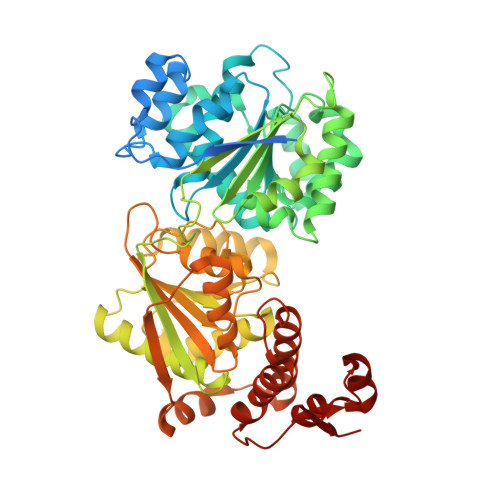
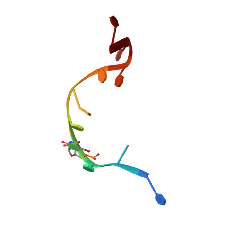
Structure of the Yeast DEAD box protein Mss116p reveals two wedges that crimp RNA

Del Campo, M.Lambowitz, A.M.

(2009) Mol Cell 35: 598-609

  • DOI: https://doi.org/10.1016/j.molcel.2009.07.032
  • Primary Citation of Related Structures:  
    3I5X, 3I5Y, 3I61, 3I62

  • PubMed Abstract: 

    The yeast DEAD box protein Mss116p is a general RNA chaperone that functions in mitochondrial group I and II intron splicing, translational activation, and RNA end processing. Here we determined high-resolution X-ray crystal structures of Mss116p complexed with an RNA oligonucleotide and ATP analogs AMP-PNP, ADP-BeF(3)(-), or ADP-AlF(4)(-). The structures show the entire helicase core acting together with a functionally important C-terminal extension. In all structures, the helicase core is in a closed conformation with a wedge alpha helix bending RNA 3' of the central bound nucleotides, as in previous DEAD box protein structures. Notably, Mss116p's C-terminal extension also bends RNA 5' of the central nucleotides, resulting in RNA crimping. Despite reported functional differences, we observe few structural changes in ternary complexes with different ATP analogs. The structures constrain models of DEAD box protein function and reveal a strand separation mechanism in which a protein uses two wedges to act as a molecular crimper.


  • Organizational Affiliation

    Institute for Cellular and Molecular Biology, University of Texas at Austin, 78712, USA.


Macromolecules

Find similar proteins by:  (by identity cutoff)  |  3D Structure
Entity ID: 1
MoleculeChains Sequence LengthOrganismDetailsImage
ATP-dependent RNA helicase MSS116563Saccharomyces cerevisiaeMutation(s): 0 
Gene Names: MSS116YD9346.05CYDR194C
EC: 3.6.1
UniProt
Find proteins for P15424 (Saccharomyces cerevisiae (strain ATCC 204508 / S288c))
Explore P15424 
Go to UniProtKB:  P15424
Entity Groups  
Sequence Clusters30% Identity50% Identity70% Identity90% Identity95% Identity100% Identity
UniProt GroupP15424
Sequence Annotations
Expand
  • Reference Sequence

Find similar nucleic acids by:  Sequence   |   3D Structure  

Entity ID: 2
MoleculeChains LengthOrganismImage
5'-R(*UP*UP*UP*(5BU)P*UP*UP*UP*UP*UP*U)-3'10N/A
Sequence Annotations
Expand
  • Reference Sequence
Small Molecules
Ligands 2 Unique
IDChains Name / Formula / InChI Key2D Diagram3D Interactions
ANP
Query on ANP

Download Ideal Coordinates CCD File 
C [auth A]PHOSPHOAMINOPHOSPHONIC ACID-ADENYLATE ESTER
C10 H17 N6 O12 P3
PVKSNHVPLWYQGJ-KQYNXXCUSA-N
MG
Query on MG

Download Ideal Coordinates CCD File 
D [auth A]MAGNESIUM ION
Mg
JLVVSXFLKOJNIY-UHFFFAOYSA-N
Experimental Data & Validation

Experimental Data

  • Method: X-RAY DIFFRACTION
  • Resolution: 2.49 Å
  • R-Value Free: 0.242 
  • R-Value Work: 0.204 
  • R-Value Observed: 0.206 
  • Space Group: P 21 21 2
Unit Cell:
Length ( Å )Angle ( ˚ )
a = 89.19α = 90
b = 126.352β = 90
c = 55.99γ = 90
Software Package:
Software NamePurpose
HKL-2000data collection
REFMACrefinement
HKL-2000data reduction
HKL-2000data scaling
REFMACphasing

Structure Validation

View Full Validation Report


Ligand Structure Quality Assessment 


Entry History 

Deposition Data

Revision History  (Full details and data files)

  • Version 1.0: 2009-09-08
    Type: Initial release
  • Version 1.1: 2011-07-13
    Changes: Advisory, Refinement description, Version format compliance
  • Version 1.2: 2023-09-06
    Changes: Data collection, Database references, Derived calculations, Refinement description