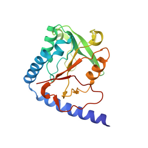
3HY6

Structure of human MTHFS with ADP


Experimental Data Snapshot

  • Method: X-RAY DIFFRACTION
  • Resolution: 2.10 Å
  • R-Value Free: 0.255 
  • R-Value Work: 0.215 
  • R-Value Observed: 0.217 

wwPDB Validation   3D Report Full Report


Ligand Structure Quality Assessment 


This is version 1.2 of the entry. See complete history


Literature

Structural basis for the inhibition of human 5,10-methenyltetrahydrofolate synthetase by N10-substituted folate analogues

Wu, D.Li, Y.Song, G.Cheng, C.Zhang, R.Joachimiak, A.Shaw, N.Liu, Z.-J.

(2009) Cancer Res 69: 7294-7301

  • DOI: https://doi.org/10.1158/0008-5472.CAN-09-1927
  • Primary Citation of Related Structures:  
    3HXT, 3HY3, 3HY4, 3HY6

  • PubMed Abstract: 

    5,10-Methenyltetrahydrofolate synthetase (MTHFS) regulates the flow of carbon through the one-carbon metabolic network, which supplies essential components for the growth and proliferation of cells. Inhibition of MTHFS in human MCF-7 breast cancer cells has been shown to arrest the growth of cells. Absence of the three-dimensional structure of human MTHFS (hMTHFS) has hampered the rational design and optimization of drug candidates. Here, we report the structures of native hMTHFS, a binary complex of hMTHFS with ADP, hMTHFS bound with the N5-iminium phosphate reaction intermediate, and an enzyme-product complex of hMTHFS. The N5-iminium phosphate captured for the first time in our crystal structure unravels a unique strategy used by hMTHFS for recognition of the substrate and provides structural basis for the regulation of enzyme activity. Binding of N10-substituted folate analogues places Y152 in the middle of the channel connecting ATP binding site with the substrate binding pocket, precluding the positioning of gamma-phosphate for a nucleophilic attack. Using the structures of hMTHFS as a guide, we have probed the role of residues surrounding the active site in catalysis by mutagenesis. The ensemble of hMTHFS structures and the mutagenesis data yield a coherent picture of the MTHFS active site, determinants of substrate specificity, and new insights into the mechanism of inhibition of hMTHFS.


  • Organizational Affiliation

    National Laboratory of Biomacromolecules, Institute of Biophysics, Chinese Academy of Sciences, Beijing, China.


Macromolecules
Find similar proteins by:  (by identity cutoff)  |  3D Structure
Entity ID: 1
MoleculeChains Sequence LengthOrganismDetailsImage
5-formyltetrahydrofolate cyclo-ligase203Homo sapiensMutation(s): 0 
Gene Names: MTHFS
EC: 6.3.3.2
UniProt & NIH Common Fund Data Resources
Find proteins for P49914 (Homo sapiens)
Explore P49914 
Go to UniProtKB:  P49914
PHAROS:  P49914
GTEx:  ENSG00000136371 
Entity Groups  
Sequence Clusters30% Identity50% Identity70% Identity90% Identity95% Identity100% Identity
UniProt GroupP49914
Sequence Annotations
Expand
  • Reference Sequence
Small Molecules
Ligands 4 Unique
IDChains Name / Formula / InChI Key2D Diagram3D Interactions
ADP
Query on ADP

Download Ideal Coordinates CCD File 
L [auth A]ADENOSINE-5'-DIPHOSPHATE
C10 H15 N5 O10 P2
XTWYTFMLZFPYCI-KQYNXXCUSA-N
PO4
Query on PO4

Download Ideal Coordinates CCD File 
I [auth A]PHOSPHATE ION
O4 P
NBIIXXVUZAFLBC-UHFFFAOYSA-K
NI
Query on NI

Download Ideal Coordinates CCD File 
B [auth A],
D [auth A],
Q [auth A]
NICKEL (II) ION
Ni
VEQPNABPJHWNSG-UHFFFAOYSA-N
MG
Query on MG

Download Ideal Coordinates CCD File 
C [auth A]
E [auth A]
F [auth A]
G [auth A]
H [auth A]
C [auth A],
E [auth A],
F [auth A],
G [auth A],
H [auth A],
J [auth A],
K [auth A],
M [auth A],
N [auth A],
O [auth A],
P [auth A],
R [auth A]
MAGNESIUM ION
Mg
JLVVSXFLKOJNIY-UHFFFAOYSA-N
Experimental Data & Validation

Experimental Data

  • Method: X-RAY DIFFRACTION
  • Resolution: 2.10 Å
  • R-Value Free: 0.255 
  • R-Value Work: 0.215 
  • R-Value Observed: 0.217 
  • Space Group: C 2 2 21
Unit Cell:
Length ( Å )Angle ( ˚ )
a = 48.511α = 90
b = 145.221β = 90
c = 59.655γ = 90
Software Package:
Software NamePurpose
HKL-2000data collection
PHASERphasing
REFMACrefinement
HKL-2000data reduction
HKL-2000data scaling

Structure Validation

View Full Validation Report


Ligand Structure Quality Assessment 


Entry History 

Deposition Data

Revision History  (Full details and data files)

  • Version 1.0: 2009-07-14
    Type: Initial release
  • Version 1.1: 2011-07-13
    Changes: Derived calculations, Version format compliance
  • Version 1.2: 2023-11-01
    Changes: Data collection, Database references, Derived calculations, Refinement description