3HC7

Crystal structure of lysin B from Mycobacteriophage D29


Experimental Data Snapshot

  • Method: X-RAY DIFFRACTION
  • Resolution: 2.00 Å
  • R-Value Free: 0.250 
  • R-Value Work: 0.205 

wwPDB Validation   3D Report Full Report


This is version 1.4 of the entry. See complete history


Literature

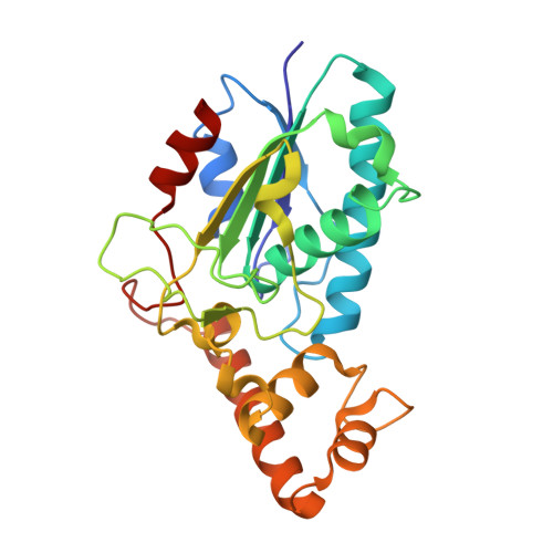
Mycobacteriophage Lysin B is a novel mycolylarabinogalactan esterase

Payne, K.Sun, Q.Sacchettini, J.Hatfull, G.F.

(2009) Mol Microbiol 73: 367-381

  • DOI: https://doi.org/10.1111/j.1365-2958.2009.06775.x
  • Primary Citation of Related Structures:  
    3HC7

  • PubMed Abstract: 

    Mycobacteriophages encounter a unique problem among phages of Gram-positive bacteria, in that lysis must not only degrade the peptidoglycan layer but also circumvent a mycolic acid-rich outer membrane covalently attached to the arabinogalactan-peptidoglycan complex. Mycobacteriophages accomplish this by producing two lysis enzymes, Lysin A (LysA) that hydrolyses peptidoglycan, and Lysin B (LysB), a novel mycolylarabinogalactan esterase, that cleaves the mycolylarabinogalactan bond to release free mycolic acids. The D29 LysB structure shows an alpha/beta hydrolase organization with a catalytic triad common to cutinases, but which contains an additional four-helix domain implicated in the binding of lipid substrates. Whereas LysA is essential for mycobacterial lysis, a Giles DeltalysB mutant mycobacteriophage is viable, but defective in the normal timing, progression and completion of host cell lysis. We propose that LysB facilitates lysis by compromising the integrity of the mycobacterial outer membrane linkage to the arabinogalactan-peptidoglycan layer.


  • Organizational Affiliation

    Department of Biological Sciences, University of Pittsburgh, Pittsburgh, PA 15260, USA.


Macromolecules
Find similar proteins by:  (by identity cutoff)  |  3D Structure
Entity ID: 1
MoleculeChains Sequence LengthOrganismDetailsImage
Gene 12 protein254Fromanvirus D29Mutation(s): 0 
Gene Names: 12gp12
UniProt
Find proteins for O64205 (Mycobacterium phage D29)
Explore O64205 
Go to UniProtKB:  O64205
Entity Groups  
Sequence Clusters30% Identity50% Identity70% Identity90% Identity95% Identity100% Identity
UniProt GroupO64205
Sequence Annotations
Expand
  • Reference Sequence
Experimental Data & Validation

Experimental Data

  • Method: X-RAY DIFFRACTION
  • Resolution: 2.00 Å
  • R-Value Free: 0.250 
  • R-Value Work: 0.205 
  • Space Group: P 43 21 2
Unit Cell:
Length ( Å )Angle ( ˚ )
a = 92.53α = 90
b = 92.53β = 90
c = 69.63γ = 90
Software Package:
Software NamePurpose
DENZOdata reduction
SCALEPACKdata scaling
SHELXphasing
PHENIXrefinement
PDB_EXTRACTdata extraction
HKL-2000data collection
SHELXDphasing

Structure Validation

View Full Validation Report


Entry History 

Deposition Data

Revision History  (Full details and data files)

  • Version 1.0: 2009-07-21
    Type: Initial release
  • Version 1.1: 2011-07-13
    Changes: Version format compliance
  • Version 1.2: 2016-03-16
    Changes: Refinement description
  • Version 1.3: 2017-11-01
    Changes: Refinement description
  • Version 1.4: 2024-02-21
    Changes: Data collection, Database references