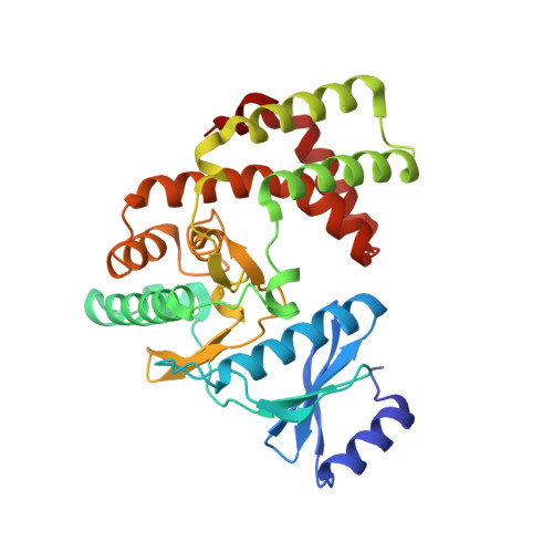
The crystal structures of substrate and nucleotide complexes of Enterococcus faecium aminoglycoside-2''-phosphotransferase-IIa [APH(2'')-IIa] provide insights into substrate selectivity in the APH(2'') subfamily.
Young, P.G., Walanj, R., Lakshmi, V., Byrnes, L.J., Metcalf, P., Baker, E.N., Vakulenko, S.B., Smith, C.A.(2009) J Bacteriol 191: 4133-4143
- PubMed: 19429619
- DOI: https://doi.org/10.1128/JB.00149-09
- Primary Citation of Related Structures:
3HAM, 3HAV - PubMed Abstract:
Aminoglycoside-2''-phosphotransferase-IIa [APH(2'')-IIa] is one of a number of homologous bacterial enzymes responsible for the deactivation of the aminoglycoside family of antibiotics and is thus a major component in bacterial resistance to these compounds. APH(2'')-IIa produces resistance to several clinically important aminoglycosides (including kanamycin and gentamicin) in both gram-positive and gram-negative bacteria, most notably in Enterococcus species. We have determined the structures of two complexes of APH(2'')-IIa, the binary gentamicin complex and a ternary complex containing adenosine-5'-(beta,gamma-methylene)triphosphate (AMPPCP) and streptomycin. This is the first crystal structure of a member of the APH(2'') family of aminoglycoside phosphotransferases. The structure of the gentamicin-APH(2'')-IIa complex was solved by multiwavelength anomalous diffraction methods from a single selenomethionine-substituted crystal and was refined to a crystallographic R factor of 0.210 (R(free), 0.271) at a resolution of 2.5 A. The structure of the AMPPCP-streptomycin complex was solved by molecular replacement using the gentamicin-APH(2'')-IIa complex as the starting model. The enzyme has a two-domain structure with the substrate binding site located in a cleft in the C-terminal domain. Gentamicin binding is facilitated by a number of conserved acidic residues lining the binding cleft, with the A and B rings of the substrate forming the majority of the interactions. The inhibitor streptomycin, although binding in the same pocket as gentamicin, is orientated such that no potential phosphorylation sites are adjacent to the catalytic aspartate residue. The binding of gentamicin and streptomycin provides structural insights into the substrate selectivity of the APH(2'') subfamily of aminoglycoside phosphotransferases, specifically, the selectivity between the 4,6-disubstituted and the 4,5-disubstituted aminoglycosides.
Organizational Affiliation:
School of Biological Sciences, University of Auckland, Auckland, New Zealand.