3H73

Crystal structure of Streptococcus pneumoniae D39 neuraminidase A precursor (NanA) in complex with DANA


Experimental Data Snapshot

  • Method: X-RAY DIFFRACTION
  • Resolution: 1.70 Å
  • R-Value Free: 0.209 
  • R-Value Work: 0.178 
  • R-Value Observed: 0.178 

wwPDB Validation   3D Report Full Report


Ligand Structure Quality Assessment 


This is version 1.3 of the entry. See complete history


Literature

Crystal structures of respiratory pathogen neuraminidases

Hsiao, Y.-S.Parker, D.Ratner, A.J.Prince, A.Tong, L.

(2009) Biochem Biophys Res Commun 380: 467-471

  • DOI: https://doi.org/10.1016/j.bbrc.2009.01.108
  • Primary Citation of Related Structures:  
    3H6J, 3H71, 3H72, 3H73

  • PubMed Abstract: 

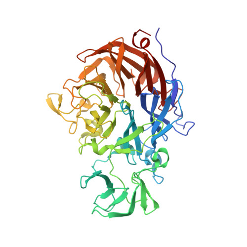
    Currently there is pressing need to develop novel therapeutic agents for the treatment of infections by the human respiratory pathogens Pseudomonas aeruginosa and Streptococcus pneumoniae. The neuraminidases of these pathogens are important for host colonization in animal models of infection and are attractive targets for drug discovery. To aid in the development of inhibitors against these neuraminidases, we have determined the crystal structures of the P. aeruginosa enzyme NanPs and S. pneumoniae enzyme NanA at 1.6 and 1.7A resolution, respectively. In situ proteolysis with trypsin was essential for the crystallization of our recombinant NanA. The active site regions of the two enzymes are strikingly different. NanA contains a deep pocket that is similar to that in canonical neuraminidases, while the NanPs active site is much more open. The comparative studies suggest that NanPs may not be a classical neuraminidase, and may have distinct natural substrates and physiological functions. This work represents an important step in the development of drugs to prevent respiratory tract colonization by these two pathogens.


  • Organizational Affiliation

    Department of Biological Sciences, Columbia University, New York, NY 10027, USA.


Macromolecules
Find similar proteins by:  (by identity cutoff)  |  3D Structure
Entity ID: 1
MoleculeChains Sequence LengthOrganismDetailsImage
Sialidase A
A, B
477Streptococcus pneumoniae R6Mutation(s): 0 
Gene Names: nanAspr1536
EC: 3.2.1.18
UniProt
Find proteins for P62576 (Streptococcus pneumoniae (strain ATCC BAA-255 / R6))
Explore P62576 
Go to UniProtKB:  P62576
Entity Groups  
Sequence Clusters30% Identity50% Identity70% Identity90% Identity95% Identity100% Identity
UniProt GroupP62576
Sequence Annotations
Expand
  • Reference Sequence
Small Molecules
Ligands 1 Unique
IDChains Name / Formula / InChI Key2D Diagram3D Interactions
DAN
Query on DAN

Download Ideal Coordinates CCD File 
C [auth A],
D [auth B]
2-DEOXY-2,3-DEHYDRO-N-ACETYL-NEURAMINIC ACID
C11 H17 N O8
JINJZWSZQKHCIP-UFGQHTETSA-N
Binding Affinity Annotations 
IDSourceBinding Affinity
DAN BindingDB:  3H73 Ki: 1600 (nM) from 1 assay(s)
IC50: min: 4800, max: 1.00e+4 (nM) from 2 assay(s)
Experimental Data & Validation

Experimental Data

  • Method: X-RAY DIFFRACTION
  • Resolution: 1.70 Å
  • R-Value Free: 0.209 
  • R-Value Work: 0.178 
  • R-Value Observed: 0.178 
  • Space Group: C 1 2 1
Unit Cell:
Length ( Å )Angle ( ˚ )
a = 158.34α = 90
b = 47.4β = 116.45
c = 137.32γ = 90
Software Package:
Software NamePurpose
ADSCdata collection
COMOphasing
CNSrefinement
DENZOdata reduction
SCALEPACKdata scaling

Structure Validation

View Full Validation Report


Ligand Structure Quality Assessment 


Entry History 

Deposition Data

Revision History  (Full details and data files)

  • Version 1.0: 2009-05-12
    Type: Initial release
  • Version 1.1: 2011-07-13
    Changes: Version format compliance
  • Version 1.2: 2020-07-29
    Type: Remediation
    Reason: Carbohydrate remediation
    Changes: Data collection, Derived calculations
  • Version 1.3: 2024-02-21
    Changes: Data collection, Database references, Structure summary