Parallel Pathways and Free-Energy Landscapes for Enzymatic Hydride Transfer Probed by Hydrostatic Pressure
Pudney, C.R., McGrory, T., Lafite, P., Pang, J., Hay, S., Leys, D., Sutcliffe, M.J., Scrutton, N.S.(2009) Chembiochem 10: 1379-1384
- PubMed: 19405065
- DOI: https://doi.org/10.1002/cbic.200900071
- Primary Citation of Related Structures:
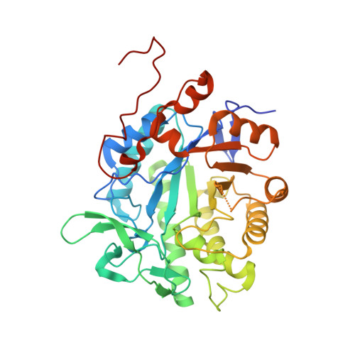
3GX9 - PubMed Abstract:
Mutation of an active-site residue in morphinone reductase leads to a conformationally rich landscape that enhances the rate of hydride transfer from NADH to FMN at standard pressure (1 bar). Increasing the pressure causes interconversion between different conformational substates in the mutant enzyme. While high pressure reduces the donor-acceptor distance in the wild-type enzyme, increased conformational freedom "dampens" its effect in the mutant.We show that hydride transfer from NADH to FMN catalysed by the N189A mutant of morphinone reductase occurs along parallel "chemical" pathways in a conformationally rich free-energy landscape. We have developed experimental kinetic and spectroscopic tools by using hydrostatic pressure to explore this free-energy landscape. The crystal structure of the N189A mutant enzyme in complex with the unreactive coenzyme analogue NADH(4) indicates that the nicotinamide moiety of the analogue is conformationally less restrained than the corresponding structure of the wild-type NADH(4) complex. This increased degree of conformational freedom in the N189A enzyme gives rise to the concept of multiple reactive configurations (MRCs), and we show that the relative population of these states across the free-energy landscape can be perturbed experimentally as a function of pressure. Specifically, the amplitudes of individual kinetic phases that were observed in stopped-flow studies of the hydride transfer reaction are sensitive to pressure; this indicates that pressure drives an altered distribution across the energy landscape. We show by absorbance spectroscopy that the loss of charge-transfer character of the enzyme-coenzyme complex is attributed to the altered population of MRCs on the landscape. The existence of a conformationally rich landscape in the N189A mutant is supported by molecular dynamics simulations at low and high pressure. The work provides firm experimental and computational support for the existence of parallel pathways arising from multiple conformational states of the enzyme-coenzyme complex. Hydrostatic pressure is a powerful and general probe of multidimensional energy landscapes that can be used to analyse experimentally parallel pathways for enzyme-catalysed reactions. We suggest that this is especially the case following directed mutation of a protein, which can lead to increased population of reactant states that are essentially inaccessible in the free-energy landscape of wild-type enzyme.
Organizational Affiliation:
Faculty of Life Sciences and Manchester Interdisciplinary Biocentre, University of Manchester, 131 Princess Street, Manchester, M1 7DN, UK.