3GV9

AmpC beta-lactamase in complex with Fragment-based Inhibitor


Experimental Data Snapshot

  • Method: X-RAY DIFFRACTION
  • Resolution: 1.80 Å
  • R-Value Free: 0.246 
  • R-Value Work: 0.210 
  • R-Value Observed: 0.212 

wwPDB Validation   3D Report Full Report


Ligand Structure Quality Assessment 


This is version 1.3 of the entry. See complete history


Literature

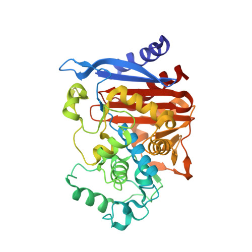
Docking for fragment inhibitors of AmpC beta-lactamase

Teotico, D.G.Babaoglu, K.Rocklin, G.J.Ferreira, R.S.Giannetti, A.M.Shoichet, B.K.

(2009) Proc Natl Acad Sci U S A 106: 7455-7460

  • DOI: https://doi.org/10.1073/pnas.0813029106
  • Primary Citation of Related Structures:  
    3GQZ, 3GR2, 3GRJ, 3GSG, 3GTC, 3GV9, 3GVB

  • PubMed Abstract: 

    Fragment screens for new ligands have had wide success, notwithstanding their constraint to libraries of 1,000-10,000 molecules. Larger libraries would be addressable were molecular docking reliable for fragment screens, but this has not been widely accepted. To investigate docking's ability to prioritize fragments, a library of >137,000 such molecules were docked against the structure of beta-lactamase. Forty-eight fragments highly ranked by docking were acquired and tested; 23 had K(i) values ranging from 0.7 to 9.2 mM. X-ray crystal structures of the enzyme-bound complexes were determined for 8 of the fragments. For 4, the correspondence between the predicted and experimental structures was high (RMSD between 1.2 and 1.4 A), whereas for another 2, the fidelity was lower but retained most key interactions (RMSD 2.4-2.6 A). Two of the 8 fragments adopted very different poses in the active site owing to enzyme conformational changes. The 48% hit rate of the fragment docking compares very favorably with "lead-like" docking and high-throughput screening against the same enzyme. To understand this, we investigated the occurrence of the fragment scaffolds among larger, lead-like molecules. Approximately 1% of commercially available fragments contain these inhibitors whereas only 10(-7)% of lead-like molecules do. This suggests that many more chemotypes and combinations of chemotypes are present among fragments than are available among lead-like molecules, contributing to the higher hit rates. The ability of docking to prioritize these fragments suggests that the technique can be used to exploit the better chemotype coverage that exists at the fragment level.


  • Organizational Affiliation

    Department of Pharmaceutical Chemistry, University of California, 1700 4th Street, MC 2550, San Francisco, CA 94158, USA.


Macromolecules
Find similar proteins by:  (by identity cutoff)  |  3D Structure
Entity ID: 1
MoleculeChains Sequence LengthOrganismDetailsImage
Beta-lactamase
A, B
358Escherichia coli K-12Mutation(s): 0 
Gene Names: ampAampCb4150JW4111
EC: 3.5.2.6
UniProt
Find proteins for P00811 (Escherichia coli (strain K12))
Explore P00811 
Go to UniProtKB:  P00811
Entity Groups  
Sequence Clusters30% Identity50% Identity70% Identity90% Identity95% Identity100% Identity
UniProt GroupP00811
Sequence Annotations
Expand
  • Reference Sequence
Small Molecules
Ligands 4 Unique
IDChains Name / Formula / InChI Key2D Diagram3D Interactions
GV9
Query on GV9

Download Ideal Coordinates CCD File 
C [auth A],
E [auth A],
F [auth A],
H [auth B]
3-(acetylamino)thiophene-2-carboxylic acid
C7 H7 N O3 S
LLKLTQJOEPWBOE-UHFFFAOYSA-N
PEG
Query on PEG

Download Ideal Coordinates CCD File 
G [auth A],
M [auth B],
O [auth B]
DI(HYDROXYETHYL)ETHER
C4 H10 O3
MTHSVFCYNBDYFN-UHFFFAOYSA-N
PO4
Query on PO4

Download Ideal Coordinates CCD File 
D [auth A],
I [auth B],
J [auth B]
PHOSPHATE ION
O4 P
NBIIXXVUZAFLBC-UHFFFAOYSA-K
DMS
Query on DMS

Download Ideal Coordinates CCD File 
K [auth B],
L [auth B],
N [auth B]
DIMETHYL SULFOXIDE
C2 H6 O S
IAZDPXIOMUYVGZ-UHFFFAOYSA-N
Binding Affinity Annotations 
IDSourceBinding Affinity
GV9 Binding MOAD:  3GV9 Ki: 7.50e+6 (nM) from 1 assay(s)
PDBBind:  3GV9 Ki: 7.50e+6 (nM) from 1 assay(s)
Experimental Data & Validation

Experimental Data

  • Method: X-RAY DIFFRACTION
  • Resolution: 1.80 Å
  • R-Value Free: 0.246 
  • R-Value Work: 0.210 
  • R-Value Observed: 0.212 
  • Space Group: C 1 2 1
Unit Cell:
Length ( Å )Angle ( ˚ )
a = 118.166α = 90
b = 78.329β = 116.59
c = 97.711γ = 90
Software Package:
Software NamePurpose
DENZOdata reduction
SCALEPACKdata scaling
REFMACrefinement
PDB_EXTRACTdata extraction

Structure Validation

View Full Validation Report


Ligand Structure Quality Assessment 


Entry History 

Deposition Data

Revision History  (Full details and data files)

  • Version 1.0: 2009-04-14
    Type: Initial release
  • Version 1.1: 2011-07-13
    Changes: Version format compliance
  • Version 1.2: 2017-11-01
    Changes: Advisory, Refinement description
  • Version 1.3: 2024-02-21
    Changes: Advisory, Data collection, Database references, Derived calculations