Crystal structures of Xanthomonas small heat shock protein provide a structural basis for an active molecular chaperone oligomer.
Hilario, E., Martin, F.J., Bertolini, M.C., Fan, L.(2011) J Mol Biol 408: 74-86
- PubMed: 21315085
- DOI: https://doi.org/10.1016/j.jmb.2011.02.004
- Primary Citation of Related Structures:
3GT6, 3GUF - PubMed Abstract:
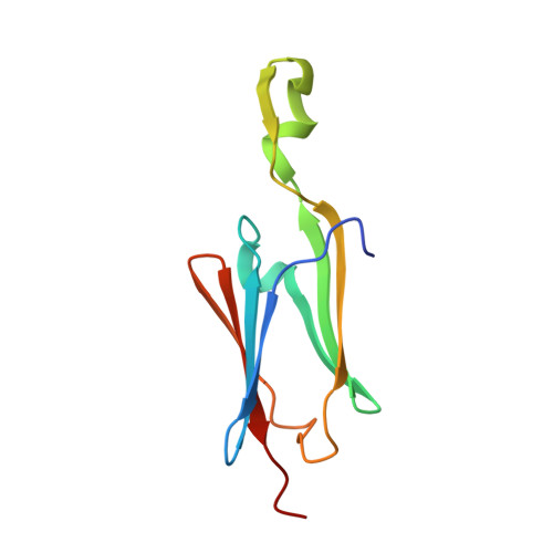
Small heat shock proteins (sHsps) are ubiquitous low-molecular-weight chaperones that prevent protein aggregation under cellular stresses. sHsps contain a structurally conserved α-crystallin domain (ACD) of about 100 amino acid residues flanked by varied N- and C-terminal extensions and usually exist as oligomers. Oligomerization is important for the biological functions of most sHsps. However, the active oligomeric states of sHsps are not defined yet. We present here crystal structures (up to 1.65 Å resolution) of the sHspA from the plant pathogen Xanthomonas (XaHspA). XaHspA forms closed or open trimers of dimers (hexamers) in crystals but exists predominantly as 36mers in solution as estimated by size-exclusion chromatography. The XaHspA monomer structures mainly consist of α-crystallin domain with disordered N- and C-terminal extensions, indicating that the extensions are flexible and not essential for the formation of dimers and 36mers. Under reducing conditions where α-lactalbumin (LA) unfolds and aggregates, XaHspA 36mers formed complexes with one LA per XaHspA dimer. Based on XaHspA dimer-dimer interactions observed in crystals, we propose that XaHspA 36mers have four possible conformations, but only XaHspA 36merB, which is formed by open hexamers in 12mer-6mer-6mer-12mer with protruding dimers accessible for substrate (unfolding protein) binding, can bind to 18 reduced LA molecules. Together, our results unravel the structural basis of an active sHsp oligomer.
Organizational Affiliation:
Department of Biochemistry, University of California, Riverside, 2482B Boyce Hall, Riverside, CA 92521-0123, USA.