3GON

Streptococcus pneumoniae Phosphomevalonate Kinase in Complex with Phosphomevalonate and AMPPNP


Experimental Data Snapshot

  • Method: X-RAY DIFFRACTION
  • Resolution: 1.90 Å
  • R-Value Free: 0.207 
  • R-Value Work: 0.185 
  • R-Value Observed: 0.185 

wwPDB Validation   3D Report Full Report


Ligand Structure Quality Assessment 


This is version 1.3 of the entry. See complete history


Literature

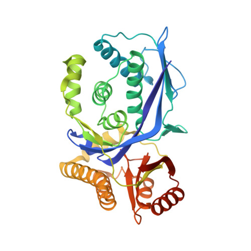
Structure of the ternary complex of phosphomevalonate kinase: the enzyme and its family

Andreassi, J.L.Vetting, M.W.Bilder, P.W.Roderick, S.L.Leyh, T.S.

(2009) Biochemistry 48: 6461-6468

  • DOI: https://doi.org/10.1021/bi900537u
  • Primary Citation of Related Structures:  
    3GON

  • PubMed Abstract: 

    The galacto-, homoserine-, mevalonate-, phosphomevalonate-kinase (GHMP) superfamily encompases a wide-range of protein function. Three members of the family (mevalonate kinase, phosphomevalonate kinase, and diphosphomevalonate decarboxylase) comprise the mevalonate pathway found in S. pneumoniae and other organisms. We have determined the 1.9 A crystal structure of phosphomevalonate kinase (PMK) from S. pneumoniae in complex with phosphomevalonate and AMPPNP.Mg(2+). Comparison of the apo and ternary PMK structures suggests that ligand binding reverses the side-chain orientations of two antiparallel lysines residues (100 and 101) with the result that Lys101 is switched into a position in which its ammonium ion is in direct contact with the beta,gamma-bridging atom of the nucleotide, where it is expected to stabilize both the ground and transition states of the reaction. Analysis of all available GHMP kinase ternary complex structures reveals that while their C(alpha)-scaffolds are highly conserved, their substrates bind in one of two conformations, which appear to be either reactive or nonreactive. The active site of PMK seems spacious enough to accommodate interconversion of the reactive and nonreactive conformers. A substantial fraction of the PMK active site is occupied by ordered water, which clusters near the charged regions of the substrate. Notably, a water pentamer that interacts extensively with the reactive groups of both substrates was discovered at the active site.


  • Organizational Affiliation

    DuPont Crop Protection, Stine-Haskell Research Center, Newark, Delaware 19711, USA.


Macromolecules
Find similar proteins by:  (by identity cutoff)  |  3D Structure
Entity ID: 1
MoleculeChains Sequence LengthOrganismDetailsImage
Phosphomevalonate kinase335Streptococcus pneumoniae R6Mutation(s): 0 
Gene Names: mvaK2spr0340
EC: 2.7.4.2
UniProt
Find proteins for Q8DR49 (Streptococcus pneumoniae (strain ATCC BAA-255 / R6))
Explore Q8DR49 
Go to UniProtKB:  Q8DR49
Entity Groups  
Sequence Clusters30% Identity50% Identity70% Identity90% Identity95% Identity100% Identity
UniProt GroupQ8DR49
Sequence Annotations
Expand
  • Reference Sequence
Small Molecules
Ligands 3 Unique
IDChains Name / Formula / InChI Key2D Diagram3D Interactions
ANP
Query on ANP

Download Ideal Coordinates CCD File 
C [auth A]PHOSPHOAMINOPHOSPHONIC ACID-ADENYLATE ESTER
C10 H17 N6 O12 P3
PVKSNHVPLWYQGJ-KQYNXXCUSA-N
PMV
Query on PMV

Download Ideal Coordinates CCD File 
B [auth A](3R)-3-HYDROXY-3-METHYL-5-(PHOSPHONOOXY)PENTANOIC ACID
C6 H13 O7 P
OKZYCXHTTZZYSK-ZCFIWIBFSA-N
MG
Query on MG

Download Ideal Coordinates CCD File 
D [auth A]MAGNESIUM ION
Mg
JLVVSXFLKOJNIY-UHFFFAOYSA-N
Experimental Data & Validation

Experimental Data

  • Method: X-RAY DIFFRACTION
  • Resolution: 1.90 Å
  • R-Value Free: 0.207 
  • R-Value Work: 0.185 
  • R-Value Observed: 0.185 
  • Space Group: P 21 21 2
Unit Cell:
Length ( Å )Angle ( ˚ )
a = 68.977α = 90
b = 115.285β = 90
c = 39.96γ = 90
Software Package:
Software NamePurpose
CNSrefinement
CTRdata collection
DENZOdata reduction
SCALEPACKdata scaling
EPMRphasing

Structure Validation

View Full Validation Report


Ligand Structure Quality Assessment 


Entry History 

Deposition Data

Revision History  (Full details and data files)

  • Version 1.0: 2009-07-21
    Type: Initial release
  • Version 1.1: 2011-07-13
    Changes: Version format compliance
  • Version 1.2: 2013-03-13
    Changes: Other
  • Version 1.3: 2023-09-06
    Changes: Data collection, Database references, Derived calculations, Refinement description