3GL9

The structure of a histidine kinase-response regulator complex sheds light into two-component signaling and reveals a novel cis autophosphorylation mechanism


Experimental Data Snapshot

  • Method: X-RAY DIFFRACTION
  • Resolution: 1.80 Å
  • R-Value Free: 0.237 
  • R-Value Work: 0.185 
  • R-Value Observed: 0.188 

wwPDB Validation   3D Report Full Report


This is version 1.2 of the entry. See complete history


Literature

Structural Insight into Partner Specificity and Phosphoryl Transfer in Two-Component Signal Transduction

Casino, P.Rubio, V.Marina, A.

(2009) Cell 139: 325-336

  • DOI: https://doi.org/10.1016/j.cell.2009.08.032
  • Primary Citation of Related Structures:  
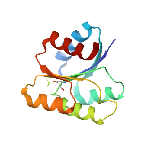
    3DGE, 3DGF, 3GL9

  • PubMed Abstract: 

    The chief mechanism used by bacteria for sensing their environment is based on two conserved proteins: a sensor histidine kinase (HK) and an effector response regulator (RR). The signal transduction process involves highly conserved domains of both proteins that mediate autokinase, phosphotransfer, and phosphatase activities whose output is a finely tuned RR phosphorylation level. Here, we report the structure of the complex between the entire cytoplasmic portion of Thermotoga maritima class I HK853 and its cognate, RR468, as well as the structure of the isolated RR468, both free and BeF(3)(-) bound. Our results provide insight into partner specificity in two-component systems, recognition of the phosphorylation state of each partner, and the catalytic mechanism of the phosphatase reaction. Biochemical analysis shows that the HK853-catalyzed autokinase reaction proceeds by a cis autophosphorylation mechanism within the HK subunit. The results suggest a model for the signal transduction mechanism in two-component systems.


  • Organizational Affiliation

    Instituto de Biomedicina de Valencia-Consejo Superior de Investigaciones Científicas (IBV-CSIC) and Centro de Investigación Biomédica en Red de Enfermedades Raras (CIBERER), Jaume Roig 11, 46010 Valencia, Spain.


Macromolecules
Find similar proteins by:  (by identity cutoff)  |  3D Structure
Entity ID: 1
MoleculeChains Sequence LengthOrganismDetailsImage
Response regulator
A, B, C, D
122Thermotoga maritimaMutation(s): 0 
UniProt
Find proteins for Q9WYT9 (Thermotoga maritima (strain ATCC 43589 / DSM 3109 / JCM 10099 / NBRC 100826 / MSB8))
Explore Q9WYT9 
Go to UniProtKB:  Q9WYT9
Entity Groups  
Sequence Clusters30% Identity50% Identity70% Identity90% Identity95% Identity100% Identity
UniProt GroupQ9WYT9
Sequence Annotations
Expand
  • Reference Sequence
Small Molecules
Modified Residues  1 Unique
IDChains TypeFormula2D DiagramParent
BFD
Query on BFD
A, B, C, D
L-PEPTIDE LINKINGC4 H6 Be F3 N O4ASP
Experimental Data & Validation

Experimental Data

  • Method: X-RAY DIFFRACTION
  • Resolution: 1.80 Å
  • R-Value Free: 0.237 
  • R-Value Work: 0.185 
  • R-Value Observed: 0.188 
  • Space Group: P 65
Unit Cell:
Length ( Å )Angle ( ˚ )
a = 155.233α = 90
b = 155.233β = 90
c = 34.482γ = 120
Software Package:
Software NamePurpose
SCALAdata scaling
PHASERphasing
REFMACrefinement
PDB_EXTRACTdata extraction
ADSCdata collection
MOSFLMdata reduction

Structure Validation

View Full Validation Report


Entry History 

Deposition Data

Revision History  (Full details and data files)

  • Version 1.0: 2009-10-27
    Type: Initial release
  • Version 1.1: 2011-07-13
    Changes: Version format compliance
  • Version 1.2: 2023-11-01
    Changes: Data collection, Database references, Derived calculations, Refinement description