3G7D

Native PhpD with Cadmium Atoms


Experimental Data Snapshot

  • Method: X-RAY DIFFRACTION
  • Resolution: 1.80 Å
  • R-Value Free: 0.246 
  • R-Value Work: 0.210 
  • R-Value Observed: 0.212 

wwPDB Validation   3D Report Full Report


This is version 1.2 of the entry. See complete history


Literature

An unusual carbon-carbon bond cleavage reaction during phosphinothricin biosynthesis.

Cicchillo, R.M.Zhang, H.Blodgett, J.A.Whitteck, J.T.Li, G.Nair, S.K.van der Donk, W.A.Metcalf, W.W.

(2009) Nature 459: 871-874

  • DOI: https://doi.org/10.1038/nature07972
  • Primary Citation of Related Structures:  
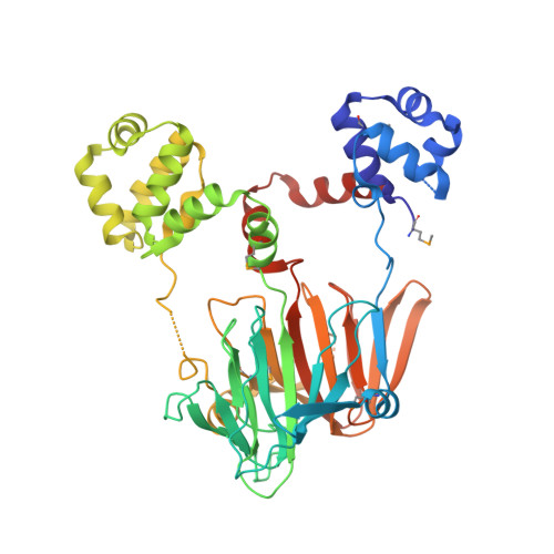
    3G7D

  • PubMed Abstract: 

    Natural products containing phosphorus-carbon bonds have found widespread use in medicine and agriculture. One such compound, phosphinothricin tripeptide, contains the unusual amino acid phosphinothricin attached to two alanine residues. Synthetic phosphinothricin (glufosinate) is a component of two top-selling herbicides (Basta and Liberty), and is widely used with resistant transgenic crops including corn, cotton and canola. Recent genetic and biochemical studies showed that during phosphinothricin tripeptide biosynthesis 2-hydroxyethylphosphonate (HEP) is converted to hydroxymethylphosphonate (HMP). Here we report the in vitro reconstitution of this unprecedented C(sp(3))-C(sp(3)) bond cleavage reaction and X-ray crystal structures of the enzyme. The protein is a mononuclear non-haem iron(ii)-dependent dioxygenase that converts HEP to HMP and formate. In contrast to most other members of this family, the oxidative consumption of HEP does not require additional cofactors or the input of exogenous electrons. The current study expands the scope of reactions catalysed by the 2-His-1-carboxylate mononuclear non-haem iron family of enzymes.


  • Organizational Affiliation

    Department of Chemistry, University of Illinois at Urbana-Champaign, Urbana, Illinois 61801, USA.


Macromolecules
Find similar proteins by:  (by identity cutoff)  |  3D Structure
Entity ID: 1
MoleculeChains Sequence LengthOrganismDetailsImage
PhpD443Streptomyces viridochromogenesMutation(s): 0 
Gene Names: Phosphinothricin biosynthetic clusterphpD
UniProt
Find proteins for Q5IW40 (Streptomyces viridochromogenes (strain DSM 40736 / JCM 4977 / BCRC 1201 / Tue 494))
Explore Q5IW40 
Go to UniProtKB:  Q5IW40
Entity Groups  
Sequence Clusters30% Identity50% Identity70% Identity90% Identity95% Identity100% Identity
UniProt GroupQ5IW40
Sequence Annotations
Expand
  • Reference Sequence
Small Molecules
Ligands 1 Unique
IDChains Name / Formula / InChI Key2D Diagram3D Interactions
CD
Query on CD

Download Ideal Coordinates CCD File 
B [auth A]
C [auth A]
D [auth A]
E [auth A]
F [auth A]
B [auth A],
C [auth A],
D [auth A],
E [auth A],
F [auth A],
G [auth A],
H [auth A],
I [auth A],
J [auth A],
K [auth A],
L [auth A],
M [auth A],
N [auth A]
CADMIUM ION
Cd
WLZRMCYVCSSEQC-UHFFFAOYSA-N
Modified Residues  1 Unique
IDChains TypeFormula2D DiagramParent
MSE
Query on MSE
A
L-PEPTIDE LINKINGC5 H11 N O2 SeMET
Experimental Data & Validation

Experimental Data

  • Method: X-RAY DIFFRACTION
  • Resolution: 1.80 Å
  • R-Value Free: 0.246 
  • R-Value Work: 0.210 
  • R-Value Observed: 0.212 
  • Space Group: C 2 2 21
Unit Cell:
Length ( Å )Angle ( ˚ )
a = 85.198α = 90
b = 95.736β = 90
c = 102.163γ = 90
Software Package:
Software NamePurpose
MAR345dtbdata collection
SHARPphasing
REFMACrefinement
HKL-2000data reduction
HKL-2000data scaling

Structure Validation

View Full Validation Report


Entry History 

Deposition Data

  • Released Date: 2009-06-09 
  • Deposition Author(s): Nair, S.K.

Revision History  (Full details and data files)

  • Version 1.0: 2009-06-09
    Type: Initial release
  • Version 1.1: 2011-07-13
    Changes: Version format compliance
  • Version 1.2: 2012-03-28
    Changes: Database references