3G5R

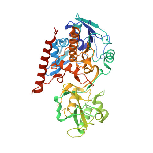
Crystal structure of Thermus thermophilus TrmFO in complex with tetrahydrofolate


Experimental Data Snapshot

  • Method: X-RAY DIFFRACTION
  • Resolution: 1.60 Å
  • R-Value Free: 0.188 
  • R-Value Work: 0.168 
  • R-Value Observed: 0.169 

wwPDB Validation   3D Report Full Report


Ligand Structure Quality Assessment 


This is version 1.3 of the entry. See complete history


Literature

Atomic structure of a folate/FAD-dependent tRNA T54 methyltransferase

Nishimasu, H.Ishitani, R.Yamashita, K.Iwashita, C.Hirata, A.Hori, H.Nureki, O.

(2009) Proc Natl Acad Sci U S A 106: 8180-8185

  • DOI: https://doi.org/10.1073/pnas.0901330106
  • Primary Citation of Related Structures:  
    3G5Q, 3G5R, 3G5S

  • PubMed Abstract: 

    tRNAs from all 3 phylogenetic domains have a 5-methyluridine at position 54 (T54) in the T-loop. The methyl group is transferred from S-adenosylmethionine by TrmA methyltransferase in most Gram-negative bacteria and some archaea and eukaryotes, whereas it is transferred from 5,10-methylenetetrahydrofolate (MTHF) by TrmFO, a folate/FAD-dependent methyltransferase, in most Gram-positive bacteria and some Gram-negative bacteria. However, the catalytic mechanism remains unclear, because the crystal structure of TrmFO has not been solved. Here, we report the crystal structures of Thermus thermophilus TrmFO in its free form, tetrahydrofolate (THF)-bound form, and glutathione-bound form at 2.1-, 1.6-, and 1.05-A resolutions, respectively. TrmFO consists of an FAD-binding domain and an insertion domain, which both share structural similarity with those of GidA, an enzyme involved in the 5-carboxymethylaminomethylation of U34 of some tRNAs. However, the overall structures of TrmFO and GidA are basically different because of their distinct domain orientations, which are consistent with their respective functional specificities. In the THF complex, the pteridin ring of THF is sandwiched between the flavin ring of FAD and the imidazole ring of a His residue. This structure provides a snapshot of the folate/FAD-dependent methyl transfer, suggesting that the transferring methylene group of MTHF is located close to the redox-active N5 atom of FAD. Furthermore, we established an in vitro system to measure the methylation activity. Our TrmFO-tRNA docking model, in combination with mutational analyses, suggests a catalytic mechanism, in which the methylene of MTHF is directly transferred onto U54, and then the exocyclic methylene of U54 is reduced by FADH(2).


  • Organizational Affiliation

    Department of Basic Medical Science, Division of Structure Biology, Institute of Medical Science, University of Tokyo, Tokyo, Japan.


Macromolecules
Find similar proteins by:  (by identity cutoff)  |  3D Structure
Entity ID: 1
MoleculeChains Sequence LengthOrganismDetailsImage
Methylenetetrahydrofolate--tRNA-(uracil-5-)-methyltransferase trmFO443Thermus thermophilus HB8Mutation(s): 0 
Gene Names: trmFOTTHA1442
EC: 2.1.1.74
UniProt
Find proteins for Q5SID2 (Thermus thermophilus (strain ATCC 27634 / DSM 579 / HB8))
Explore Q5SID2 
Go to UniProtKB:  Q5SID2
Entity Groups  
Sequence Clusters30% Identity50% Identity70% Identity90% Identity95% Identity100% Identity
UniProt GroupQ5SID2
Sequence Annotations
Expand
  • Reference Sequence
Small Molecules
Ligands 4 Unique
IDChains Name / Formula / InChI Key2D Diagram3D Interactions
FAD
Query on FAD

Download Ideal Coordinates CCD File 
B [auth A]FLAVIN-ADENINE DINUCLEOTIDE
C27 H33 N9 O15 P2
VWWQXMAJTJZDQX-UYBVJOGSSA-N
THG
Query on THG

Download Ideal Coordinates CCD File 
C [auth A](6S)-5,6,7,8-TETRAHYDROFOLATE
C19 H23 N7 O6
MSTNYGQPCMXVAQ-RYUDHWBXSA-N
EDO
Query on EDO

Download Ideal Coordinates CCD File 
E [auth A]
F [auth A]
G [auth A]
H [auth A]
I [auth A]
E [auth A],
F [auth A],
G [auth A],
H [auth A],
I [auth A],
J [auth A]
1,2-ETHANEDIOL
C2 H6 O2
LYCAIKOWRPUZTN-UHFFFAOYSA-N
CA
Query on CA

Download Ideal Coordinates CCD File 
D [auth A]CALCIUM ION
Ca
BHPQYMZQTOCNFJ-UHFFFAOYSA-N
Experimental Data & Validation

Experimental Data

  • Method: X-RAY DIFFRACTION
  • Resolution: 1.60 Å
  • R-Value Free: 0.188 
  • R-Value Work: 0.168 
  • R-Value Observed: 0.169 
  • Space Group: P 1 21 1
Unit Cell:
Length ( Å )Angle ( ˚ )
a = 46.809α = 90
b = 97.023β = 101.29
c = 53.794γ = 90
Software Package:
Software NamePurpose
HKL-2000data collection
MOLREPphasing
PHENIXrefinement
HKL-2000data reduction
HKL-2000data scaling

Structure Validation

View Full Validation Report


Ligand Structure Quality Assessment 


Entry History 

Deposition Data

Revision History  (Full details and data files)

  • Version 1.0: 2009-05-19
    Type: Initial release
  • Version 1.1: 2011-07-13
    Changes: Version format compliance
  • Version 1.2: 2011-08-24
    Changes: Non-polymer description
  • Version 1.3: 2023-11-01
    Changes: Data collection, Database references, Derived calculations, Refinement description