3G4F

Crystal Structure of (+)- -Cadinene Synthase from Gossypium arboreum in complex with 2-fluorofarnesyl diphosphate


Experimental Data Snapshot

  • Method: X-RAY DIFFRACTION
  • Resolution: 2.65 Å
  • R-Value Free: 0.256 
  • R-Value Work: 0.201 
  • R-Value Observed: 0.206 

wwPDB Validation   3D Report Full Report


Ligand Structure Quality Assessment 


This is version 1.2 of the entry. See complete history


Literature

Crystal structure of (+)-delta-cadinene synthase from Gossypium arboreum and evolutionary divergence of metal binding motifs for catalysis.

Gennadios, H.A.Gonzalez, V.Di Costanzo, L.Li, A.Yu, F.Miller, D.J.Allemann, R.K.Christianson, D.W.

(2009) Biochemistry 48: 6175-6183

  • DOI: https://doi.org/10.1021/bi900483b
  • Primary Citation of Related Structures:  
    3G4D, 3G4F

  • PubMed Abstract: 

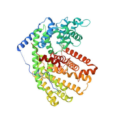
    (+)-Delta-cadinene synthase (DCS) from Gossypium arboreum (tree cotton) is a sesquiterpene cyclase that catalyzes the cyclization of farnesyl diphosphate in the first committed step of the biosynthesis of gossypol, a phytoalexin that defends the plant from bacterial and fungal pathogens. Here, we report the X-ray crystal structure of unliganded DCS at 2.4 A resolution and the structure of its complex with three putative Mg(2+) ions and the substrate analogue inhibitor 2-fluorofarnesyl diphosphate (2F-FPP) at 2.75 A resolution. These structures illuminate unusual features that accommodate the trinuclear metal cluster required for substrate binding and catalysis. Like other terpenoid cyclases, DCS contains a characteristic aspartate-rich D(307)DTYD(311) motif on helix D that interacts with Mg(2+)(A) and Mg(2+)(C). However, DCS appears to be unique among terpenoid cyclases in that it does not contain the "NSE/DTE" motif on helix H that specifically chelates Mg(2+)(B), which is usually found as the signature sequence (N,D)D(L,I,V)X(S,T)XXXE (boldface indicates Mg(2+)(B) ligands). Instead, DCS contains a second aspartate-rich motif, D(451)DVAE(455), that interacts with Mg(2+)(B). In this regard, DCS is more similar to the isoprenoid chain elongation enzyme farnesyl diphosphate synthase, which also contains two aspartate-rich motifs, rather than the greater family of terpenoid cyclases. Nevertheless, the structure of the DCS-2F-FPP complex shows that the structure of the trinuclear magnesium cluster is generally similar to that of other terpenoid cyclases despite the alternative Mg(2+)(B) binding motif. Analyses of DCS mutants with alanine substitutions in the D(307)DTYD(311) and D(451)DVAE(455) segments reveal the contributions of these segments to catalysis.


  • Organizational Affiliation

    Roy and Diana Vagelos Laboratories, Department of Chemistry, University of Pennsylvania, Philadelphia, Pennsylvania 19104-6323, USA.


Macromolecules
Find similar proteins by:  (by identity cutoff)  |  3D Structure
Entity ID: 1
MoleculeChains Sequence LengthOrganismDetailsImage
(+)-delta-cadinene synthase isozyme XC1
A, B
554Gossypium arboreumMutation(s): 0 
EC: 4.2.3.13
UniProt
Find proteins for Q39761 (Gossypium arboreum)
Explore Q39761 
Go to UniProtKB:  Q39761
Entity Groups  
Sequence Clusters30% Identity50% Identity70% Identity90% Identity95% Identity100% Identity
UniProt GroupQ39761
Sequence Annotations
Expand
  • Reference Sequence
Small Molecules
Ligands 3 Unique
IDChains Name / Formula / InChI Key2D Diagram3D Interactions
FPF
Query on FPF

Download Ideal Coordinates CCD File 
E [auth A],
I [auth B]
(2Z,6E)-2-fluoro-3,7,11-trimethyldodeca-2,6,10-trien-1-yl trihydrogen diphosphate
C15 H27 F O7 P2
DXHJZCKJWKWTCH-WISUYLHISA-N
BME
Query on BME

Download Ideal Coordinates CCD File 
C [auth A],
D [auth A]
BETA-MERCAPTOETHANOL
C2 H6 O S
DGVVWUTYPXICAM-UHFFFAOYSA-N
MG
Query on MG

Download Ideal Coordinates CCD File 
F [auth A]
G [auth A]
H [auth A]
J [auth B]
K [auth B]
F [auth A],
G [auth A],
H [auth A],
J [auth B],
K [auth B],
L [auth B]
MAGNESIUM ION
Mg
JLVVSXFLKOJNIY-UHFFFAOYSA-N
Binding Affinity Annotations 
IDSourceBinding Affinity
FPF PDBBind:  3G4F IC50: 3.00e+4 (nM) from 1 assay(s)
Experimental Data & Validation

Experimental Data

  • Method: X-RAY DIFFRACTION
  • Resolution: 2.65 Å
  • R-Value Free: 0.256 
  • R-Value Work: 0.201 
  • R-Value Observed: 0.206 
  • Space Group: P 21 3
Unit Cell:
Length ( Å )Angle ( ˚ )
a = 158.195α = 90
b = 158.195β = 90
c = 158.195γ = 90
Software Package:
Software NamePurpose
ADSCdata collection
PHASERphasing
PHENIXrefinement
HKL-2000data reduction
SCALEPACKdata scaling

Structure Validation

View Full Validation Report


Ligand Structure Quality Assessment 


Entry History 

Deposition Data

Revision History  (Full details and data files)

  • Version 1.0: 2009-06-23
    Type: Initial release
  • Version 1.1: 2011-07-13
    Changes: Version format compliance
  • Version 1.2: 2024-04-03
    Changes: Data collection, Database references, Derived calculations, Refinement description