3FV3

Secreted aspartic protease 1 from Candida parapsilosis in complex with pepstatin A


Experimental Data Snapshot

  • Method: X-RAY DIFFRACTION
  • Resolution: 1.85 Å
  • R-Value Free: 0.190 
  • R-Value Work: 0.166 
  • R-Value Observed: 0.166 

wwPDB Validation   3D Report Full Report


This is version 1.4 of the entry. See complete history


Literature

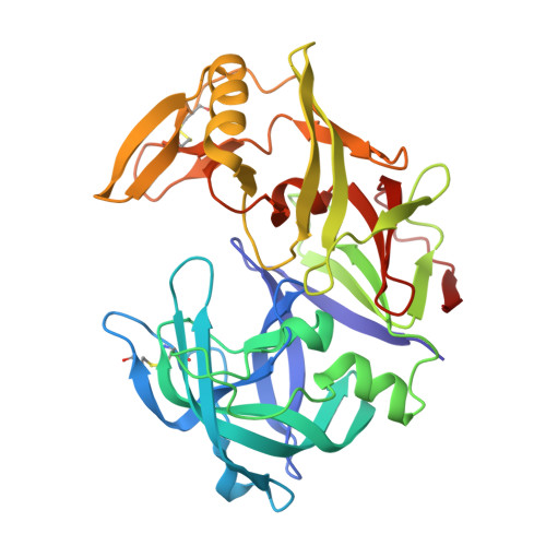
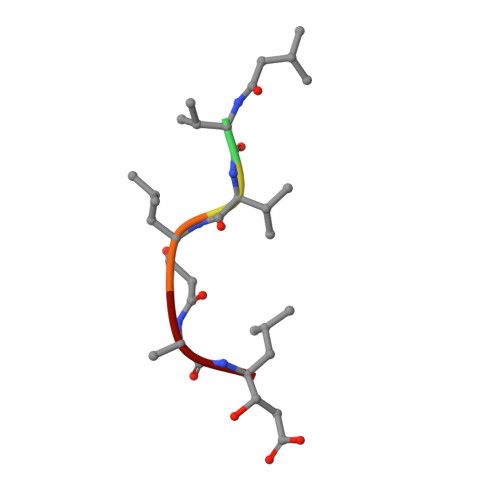
The crystal structure of the secreted aspartic protease 1 from Candida parapsilosis in complex with pepstatin A

Dostal, J.Brynda, J.Hruskova-Heidingsfeldova, O.Sieglova, I.Pichova, I.Rezacova, P.

(2009) J Struct Biol 167: 145-152

  • DOI: https://doi.org/10.1016/j.jsb.2009.04.004
  • Primary Citation of Related Structures:  
    3FV3

  • PubMed Abstract: 

    Opportunistic pathogens of the genus Candida cause infections representing a major threat to long-term survival of immunocompromised patients. Virulence of the Candida pathogens is enhanced by production of extracellular proteolytic enzymes and secreted aspartic proteases (Saps) are therefore studied as potential virulence factors and possible targets for therapeutic drug design. Candida parapsilosis is less invasive than C. albicans, however, it is one of the leading causative agents of yeast infections. We report three-dimensional crystal structure of Sapp1p from C. parapsilosis in complex with pepstatin A, the classical inhibitor of aspartic proteases. The structure of Sapp1p was determined from protein isolated from its natural source and represents the first structure of Sap from C. parapsilosis. Overall fold and topology of Sapp1p is very similar to the archetypic fold of monomeric aspartic protease family and known structures of Sap isoenzymes from C. albicans and Sapt1p from C. tropicalis. Structural comparison revealed noticeable differences in the structure of loops surrounding the active site. This resulted in differential character, shape, and size of the substrate binding site explaining divergent substrate specificities and inhibitor affinities. Determination of structures of Sap isoenzymes from various species might contribute to the development of new Sap-specific inhibitors.


  • Organizational Affiliation

    Gilead Sciences and IOCB Research Centre, Prague, Czech Republic.


Macromolecules
Find similar proteins by:  (by identity cutoff)  |  3D Structure
Entity ID: 1
MoleculeChains Sequence LengthOrganismDetailsImage
Sapp1p-secreted aspartic protease 1339Candida parapsilosisMutation(s): 0 
EC: 3.4.23.24
UniProt
Find proteins for P32951 (Candida parapsilosis)
Explore P32951 
Go to UniProtKB:  P32951
Entity Groups  
Sequence Clusters30% Identity50% Identity70% Identity90% Identity95% Identity100% Identity
UniProt GroupP32951
Sequence Annotations
Expand
  • Reference Sequence

Find similar proteins by:  Sequence   |   3D Structure  

Entity ID: 2
MoleculeChains Sequence LengthOrganismDetailsImage
pepstatin A6Streptomyces argenteolus subsp. toyonakensisMutation(s): 0 
Sequence Annotations
Expand
  • Reference Sequence
Small Molecules
Ligands 2 Unique
IDChains Name / Formula / InChI Key2D Diagram3D Interactions
SO4
Query on SO4

Download Ideal Coordinates CCD File 
CA [auth D]
DA [auth D]
HA [auth E]
IA [auth E]
NA [auth F]
CA [auth D],
DA [auth D],
HA [auth E],
IA [auth E],
NA [auth F],
OA [auth F],
Q [auth A],
QA [auth G],
RA [auth G],
SA [auth G],
TA [auth G],
U [auth B],
UA [auth G],
XA [auth H],
YA [auth H]
SULFATE ION
O4 S
QAOWNCQODCNURD-UHFFFAOYSA-L
GOL
Query on GOL

Download Ideal Coordinates CCD File 
AA [auth C]
AB [auth H]
BA [auth C]
BB [auth H]
CB [auth H]
AA [auth C],
AB [auth H],
BA [auth C],
BB [auth H],
CB [auth H],
DB [auth H],
EA [auth D],
FA [auth D],
GA [auth D],
JA [auth E],
KA [auth E],
LA [auth E],
MA [auth E],
PA [auth F],
R [auth A],
S [auth A],
T [auth A],
V [auth B],
VA [auth G],
W [auth B],
WA [auth G],
X [auth B],
Y [auth B],
Z [auth C],
ZA [auth H]
GLYCEROL
C3 H8 O3
PEDCQBHIVMGVHV-UHFFFAOYSA-N
Experimental Data & Validation

Experimental Data

  • Method: X-RAY DIFFRACTION
  • Resolution: 1.85 Å
  • R-Value Free: 0.190 
  • R-Value Work: 0.166 
  • R-Value Observed: 0.166 
  • Space Group: P 1 21 1
Unit Cell:
Length ( Å )Angle ( ˚ )
a = 86.488α = 90
b = 194.247β = 91.52
c = 97.147γ = 90
Software Package:
Software NamePurpose
HKL-2000data collection
MOLREPphasing
REFMACrefinement
HKL-3000data reduction
HKL-3000data scaling

Structure Validation

View Full Validation Report


Entry History 

Deposition Data

Revision History  (Full details and data files)

  • Version 1.0: 2009-05-19
    Type: Initial release
  • Version 1.1: 2011-07-13
    Changes: Advisory, Version format compliance
  • Version 1.2: 2013-02-27
    Changes: Other
  • Version 1.3: 2023-11-01
    Changes: Data collection, Database references, Derived calculations, Refinement description
  • Version 1.4: 2023-11-22
    Changes: Data collection