3FOP

A Triangular Cytochrome b562 Superstructure Mediated by Ni Coordination - Hexagonal Form


Experimental Data Snapshot

  • Method: X-RAY DIFFRACTION
  • Resolution: 3.00 Å
  • R-Value Free: 0.277 
  • R-Value Work: 0.221 
  • R-Value Observed: 0.225 

wwPDB Validation   3D Report Full Report


Ligand Structure Quality Assessment 


This is version 1.3 of the entry. See complete history


Literature

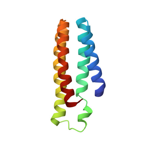
A superprotein triangle driven by nickel(II) coordination: exploiting non-natural metal ligands in protein self-assembly.

Radford, R.J.Tezcan, F.A.

(2009) J Am Chem Soc 131: 9136-9137

  • DOI: https://doi.org/10.1021/ja9000695
  • Primary Citation of Related Structures:  
    3FOO, 3FOP

  • PubMed Abstract: 

    We previously devised a strategy (metal-directed protein self-assembly, MDPSA) that utilizes the simultaneous stability, lability, and directionality of metal-ligand bonds to drive protein-protein interactions. Here we show that both the structural and functional scopes of MDPSA can be broadened by incorporation of non-natural metal-chelating ligands onto protein surfaces. A cytochrome cb(562) variant, MBP-Phen1, which features a covalently attached phenanthroline (Phen) group on its surface, self-assembles into an unusual triangular architecture (Ni(3):MBP-Phen1(3)) upon binding Ni as a result of specific Phen-protein interactions. The crystal structure of Ni(3):MBP-Phen1(3) reveals that the Phen group is buried in a small pocket on the protein surface, which results in an unsaturated Ni coordination environment.


  • Organizational Affiliation

    Department of Chemistry and Biochemistry, University of California, San Diego, 9500 Gilman Drive, La Jolla, California 92093-0356, USA.


Macromolecules
Find similar proteins by:  (by identity cutoff)  |  3D Structure
Entity ID: 1
MoleculeChains Sequence LengthOrganismDetailsImage
Soluble cytochrome b562
A, B
106Escherichia coliMutation(s): 7 
Gene Names: cybC
UniProt
Find proteins for P0ABE7 (Escherichia coli)
Explore P0ABE7 
Go to UniProtKB:  P0ABE7
Entity Groups  
Sequence Clusters30% Identity50% Identity70% Identity90% Identity95% Identity100% Identity
UniProt GroupP0ABE7
Sequence Annotations
Expand
  • Reference Sequence
Small Molecules
Ligands 3 Unique
IDChains Name / Formula / InChI Key2D Diagram3D Interactions
HEM
Query on HEM

Download Ideal Coordinates CCD File 
C [auth A],
G [auth B]
PROTOPORPHYRIN IX CONTAINING FE
C34 H32 Fe N4 O4
KABFMIBPWCXCRK-RGGAHWMASA-L
PXX
Query on PXX

Download Ideal Coordinates CCD File 
D [auth A],
H [auth B]
N-1,10-phenanthrolin-5-ylacetamide
C14 H11 N3 O
AAJXINSCZMZERD-UHFFFAOYSA-N
NI
Query on NI

Download Ideal Coordinates CCD File 
E [auth A],
F [auth A],
I [auth B],
J [auth B]
NICKEL (II) ION
Ni
VEQPNABPJHWNSG-UHFFFAOYSA-N
Experimental Data & Validation

Experimental Data

  • Method: X-RAY DIFFRACTION
  • Resolution: 3.00 Å
  • R-Value Free: 0.277 
  • R-Value Work: 0.221 
  • R-Value Observed: 0.225 
  • Space Group: P 63 2 2
Unit Cell:
Length ( Å )Angle ( ˚ )
a = 104.897α = 90
b = 104.897β = 90
c = 107.925γ = 120
Software Package:
Software NamePurpose
MOSFLMdata reduction
SCALAdata scaling
MOLREPphasing
REFMACrefinement
PDB_EXTRACTdata extraction

Structure Validation

View Full Validation Report


Ligand Structure Quality Assessment 


Entry History 

Deposition Data

Revision History  (Full details and data files)

  • Version 1.0: 2009-07-14
    Type: Initial release
  • Version 1.1: 2011-07-13
    Changes: Version format compliance
  • Version 1.2: 2021-10-20
    Changes: Database references, Derived calculations
  • Version 1.3: 2023-09-06
    Changes: Data collection, Refinement description