


Structural and Site-directed Mutagenesis Study of Versatile Peroxidase Oxidizing both Mn(II) and Aromatic Substrates
Piontek, K., Choinowski, T., Perez-Boada, M., Ruiz-Duenas, F.J., Martinez, M.J., Plattner, D.A., Martinez, A.T.To be published.
Experimental Data Snapshot
Entity ID: 1 | |||||
|---|---|---|---|---|---|
| Molecule | Chains | Sequence Length | Organism | Details | Image |
| Versatile peroxidase VPL2 | 331 | Pleurotus eryngii | Mutation(s): 0 Gene Names: vpl2 EC: 1.11.1.16 | 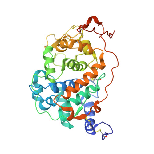 | |
UniProt | |||||
Find proteins for O94753 (Pleurotus eryngii) Explore O94753 Go to UniProtKB: O94753 | |||||
Entity Groups | |||||
| Sequence Clusters | 30% Identity50% Identity70% Identity90% Identity95% Identity100% Identity | ||||
| UniProt Group | O94753 | ||||
Sequence AnnotationsExpand | |||||
| |||||
| Ligands 5 Unique | |||||
|---|---|---|---|---|---|
| ID | Chains | Name / Formula / InChI Key | 2D Diagram | 3D Interactions | |
| HEM Query on HEM | B [auth A] | PROTOPORPHYRIN IX CONTAINING FE C34 H32 Fe N4 O4 KABFMIBPWCXCRK-RGGAHWMASA-L |  | ||
| CAC Query on CAC | O [auth A], P [auth A] | CACODYLATE ION C2 H6 As O2 OGGXGZAMXPVRFZ-UHFFFAOYSA-M |  | ||
| ZN Query on ZN | E [auth A] F [auth A] G [auth A] I [auth A] J [auth A] | ZINC ION Zn PTFCDOFLOPIGGS-UHFFFAOYSA-N |  | ||
| FE Query on FE | H [auth A], M [auth A], N [auth A] | FE (III) ION Fe VTLYFUHAOXGGBS-UHFFFAOYSA-N |  | ||
| CA Query on CA | C [auth A], D [auth A] | CALCIUM ION Ca BHPQYMZQTOCNFJ-UHFFFAOYSA-N |  | ||
| Length ( Å ) | Angle ( ˚ ) |
|---|---|
| a = 62.683 | α = 90 |
| b = 62.683 | β = 90 |
| c = 98.217 | γ = 90 |
| Software Name | Purpose |
|---|---|
| MAR345dtb | data collection |
| AMoRE | phasing |
| REFMAC | refinement |
| DENZO | data reduction |
| SCALEPACK | data scaling |
RCSB PDB (citation) is hosted by
RCSB PDB is a member of the

