3FHK

Crystal structure of APC1446, B.subtilis YphP disulfide isomerase


Experimental Data Snapshot

  • Method: X-RAY DIFFRACTION
  • Resolution: 2.30 Å
  • R-Value Free: 0.246 
  • R-Value Work: 0.190 
  • R-Value Observed: 0.193 

wwPDB Validation   3D Report Full Report


This is version 1.4 of the entry. See complete history


Literature

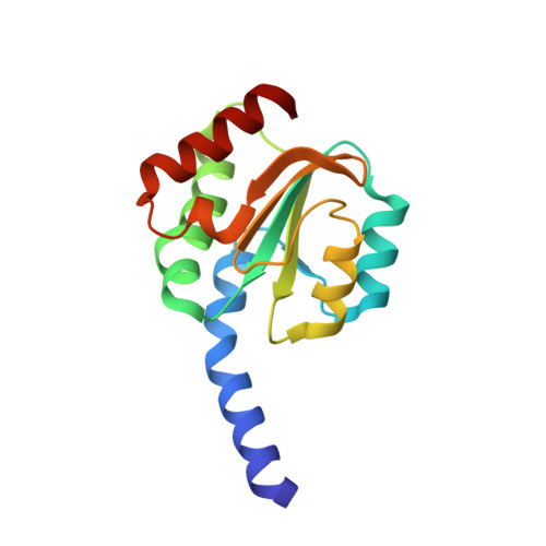
Structure and function of Bacillus subtilis YphP, a prokaryotic disulfide isomerase with a CXC catalytic motif .

Derewenda, U.Boczek, T.Gorres, K.L.Yu, M.Hung, L.W.Cooper, D.Joachimiak, A.Raines, R.T.Derewenda, Z.S.

(2009) Biochemistry 48: 8664-8671

  • DOI: https://doi.org/10.1021/bi900437z
  • Primary Citation of Related Structures:  
    3FHK

  • PubMed Abstract: 

    The DUF1094 family contains over 100 bacterial proteins, all containing a conserved CXC motif, with unknown function. We solved the crystal structure of the Bacillus subtilis representative, the product of the yphP gene. The protein shows remarkable structural similarity to thioredoxins, with a canonical alphabetaalphabetaalphabetabetaalpha topology, despite low amino acid sequence identity to thioredoxin. The CXC motif is found in the loop immediately downstream of the first beta-strand, in a location equivalent to the CXXC motif of thioredoxins, with the first Cys occupying a position equivalent to the first Cys in canonical thioredoxin. The experimentally determined reduction potential of YphP is E degrees' = -130 mV, significantly higher than that of thioredoxin and consistent with disulfide isomerase activity. Functional assays confirmed that the protein displays a level of isomerase activity that might be biologically significant. We propose a mechanism by which the members of this family catalyze isomerization using the CXC catalytic site.


  • Organizational Affiliation

    Department of Molecular Physiology and Biological Physics and the ISFI PSI2 Center, University of Virginia School of Medicine, Charlottesville, Virginia 22908-0736, USA.


Macromolecules
Find similar proteins by:  (by identity cutoff)  |  3D Structure
Entity ID: 1
MoleculeChains Sequence LengthOrganismDetailsImage
UPF0403 protein yphPA,
B [auth D],
C [auth E],
D [auth F]
147Bacillus subtilisMutation(s): 2 
Gene Names: BSU21860yphP
UniProt
Find proteins for P54170 (Bacillus subtilis (strain 168))
Explore P54170 
Go to UniProtKB:  P54170
Entity Groups  
Sequence Clusters30% Identity50% Identity70% Identity90% Identity95% Identity100% Identity
UniProt GroupP54170
Sequence Annotations
Expand
  • Reference Sequence
Small Molecules
Ligands 1 Unique
IDChains Name / Formula / InChI Key2D Diagram3D Interactions
SO4
Query on SO4

Download Ideal Coordinates CCD File 
E [auth A]
F [auth A]
G [auth A]
H [auth D]
I [auth D]
E [auth A],
F [auth A],
G [auth A],
H [auth D],
I [auth D],
J [auth D],
K [auth E],
L [auth E],
M [auth E],
N [auth F],
O [auth F],
P [auth F]
SULFATE ION
O4 S
QAOWNCQODCNURD-UHFFFAOYSA-L
Experimental Data & Validation

Experimental Data

  • Method: X-RAY DIFFRACTION
  • Resolution: 2.30 Å
  • R-Value Free: 0.246 
  • R-Value Work: 0.190 
  • R-Value Observed: 0.193 
  • Space Group: P 41 21 2
Unit Cell:
Length ( Å )Angle ( ˚ )
a = 68.161α = 90
b = 68.161β = 90
c = 302.024γ = 90
Software Package:
Software NamePurpose
PHENIXrefinement
REFMACrefinement
HKL-2000data reduction
HKL-2000data scaling
SHELXEmodel building

Structure Validation

View Full Validation Report


Entry History 

Revision History  (Full details and data files)

  • Version 1.0: 2009-09-01
    Type: Initial release
  • Version 1.1: 2011-07-13
    Changes: Version format compliance
  • Version 1.2: 2019-07-24
    Changes: Data collection, Refinement description
  • Version 1.3: 2021-10-20
    Changes: Database references, Derived calculations
  • Version 1.4: 2024-02-21
    Changes: Data collection