3FDQ

Recognition of AT-rich DNA binding sites by the MogR Repressor


Experimental Data Snapshot

  • Method: X-RAY DIFFRACTION
  • Resolution: 1.75 Å
  • R-Value Free: 0.232 
  • R-Value Work: 0.217 
  • R-Value Observed: 0.217 

wwPDB Validation   3D Report Full Report


This is version 1.2 of the entry. See complete history


Literature

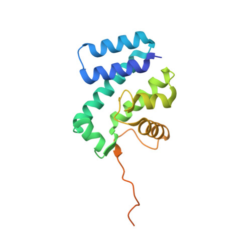
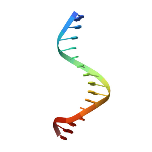
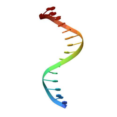
Recognition of AT-Rich DNA Binding Sites by the MogR Repressor.

Shen, A.Higgins, D.E.Panne, D.

(2009) Structure 17: 769-777

  • DOI: https://doi.org/10.1016/j.str.2009.02.018
  • Primary Citation of Related Structures:  
    3FDQ

  • PubMed Abstract: 

    The MogR transcriptional repressor of the intracellular pathogen Listeria monocytogenes recognizes AT-rich binding sites in promoters of flagellar genes to downregulate flagellar gene expression during infection. We describe here the 1.8 A resolution crystal structure of MogR bound to the recognition sequence 5' ATTTTTTAAAAAAAT 3' present within the flaA promoter region. Our structure shows that MogR binds as a dimer. Each half-site is recognized in the major groove by a helix-turn-helix motif and in the minor groove by a loop from the symmetry-related molecule, resulting in a "crossover" binding mode. This oversampling through minor groove interactions is important for specificity. The MogR binding site has structural features of A-tract DNA and is bent by approximately 52 degrees away from the dimer. The structure explains how MogR achieves binding specificity in the AT-rich genome of L. monocytogenes and explains the evolutionary conservation of A-tract sequence elements within promoter regions of MogR-regulated flagellar genes.


  • Organizational Affiliation

    Department of Microbiology and Molecular Genetics, Harvard Medical School, Boston, MA 02115, USA.


Macromolecules

Find similar proteins by:  (by identity cutoff)  |  3D Structure
Entity ID: 1
MoleculeChains Sequence LengthOrganismDetailsImage
Motility gene repressor mogR
A, B
170Listeria monocytogenesMutation(s): 0 
Gene Names: lmo0674mogR
UniProt
Find proteins for P0DJO8 (Listeria monocytogenes serovar 1/2a (strain ATCC BAA-679 / EGD-e))
Explore P0DJO8 
Go to UniProtKB:  P0DJO8
Entity Groups  
Sequence Clusters30% Identity50% Identity70% Identity90% Identity95% Identity100% Identity
UniProt GroupP0DJO8
Sequence Annotations
Expand
  • Reference Sequence

Find similar nucleic acids by:  Sequence   |   3D Structure  

Entity ID: 2
MoleculeChains LengthOrganismImage
5'-D(*AP*TP*TP*TP*TP*TP*TP*AP*AP*AP*AP*AP*AP*AP*T)-3'15N/A
Sequence Annotations
Expand
  • Reference Sequence

Find similar nucleic acids by:  Sequence   |   3D Structure  

Entity ID: 3
MoleculeChains LengthOrganismImage
5'-D(*TP*AP*TP*TP*TP*TP*TP*TP*TP*AP*AP*AP*AP*AP*A)-3'15N/A
Sequence Annotations
Expand
  • Reference Sequence
Experimental Data & Validation

Experimental Data

  • Method: X-RAY DIFFRACTION
  • Resolution: 1.75 Å
  • R-Value Free: 0.232 
  • R-Value Work: 0.217 
  • R-Value Observed: 0.217 
  • Space Group: P 1 21 1
Unit Cell:
Length ( Å )Angle ( ˚ )
a = 48.344α = 90
b = 93.597β = 113.41
c = 60.768γ = 90
Software Package:
Software NamePurpose
ADSCdata collection
SHELXDphasing
CNSrefinement
HKL-2000data reduction
HKL-2000data scaling

Structure Validation

View Full Validation Report


Entry History 

Deposition Data

Revision History  (Full details and data files)

  • Version 1.0: 2009-06-02
    Type: Initial release
  • Version 1.1: 2011-07-13
    Changes: Version format compliance
  • Version 1.2: 2023-12-27
    Changes: Data collection, Database references