3FB4

Crystal structure of adenylate kinase from Marinibacillus marinus


Experimental Data Snapshot

  • Method: X-RAY DIFFRACTION
  • Resolution: 2.00 Å
  • R-Value Free: 0.226 
  • R-Value Work: 0.180 
  • R-Value Observed: 0.183 

wwPDB Validation   3D Report Full Report


Ligand Structure Quality Assessment 


This is version 1.2 of the entry. See complete history


Literature

Structure and biochemical characterization of an adenylate kinase originating from the psychrophilic organism Marinibacillus marinus.

Davlieva, M.Shamoo, Y.

(2009) Acta Crystallogr Sect F Struct Biol Cryst Commun 65: 751-756

  • DOI: https://doi.org/10.1107/S1744309109024348
  • Primary Citation of Related Structures:  
    3FB4

  • PubMed Abstract: 

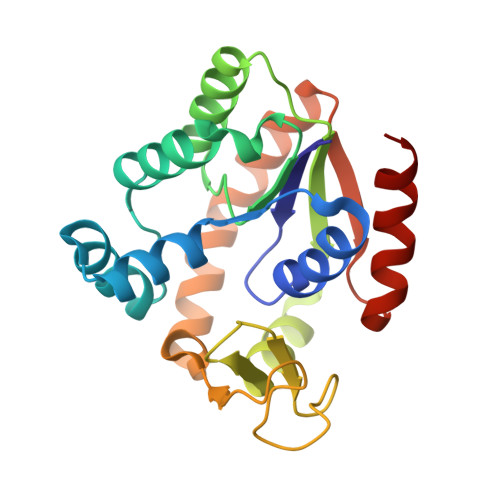
    Adenylate kinases (AKs; EC 2.7.4.3) are essential members of the NMP kinase family that maintain cellular homeostasis by the interconversion of AMP, ADP and ATP. AKs play a critical role in adenylate homeostasis across all domains of life and have been used extensively as prototypes for the study of protein adaptation and the relationship of protein dynamics and stability to function. To date, kinetic studies of psychrophilic AKs have not been performed. In order to broaden understanding of extremophilic adaptation, the kinetic parameters of adenylate kinase from the psychrophile Marinibacillus marinus were examined and the crystal structure of this cold-adapted enzyme was determined at 2.0 A resolution. As expected, the overall structure and topology of the psychrophilic M. marinus AK are similar to those of mesophilic and thermophilic AKs. The thermal denaturation midpoint of M. marinus AK (321.1 K) is much closer to that of the mesophile Bacillus subtilis (320.7 K) than the more closely related psychrophile B. globisporus (316.4 K). In addition, the enzymatic properties of M. marinus AK are quite close to those of the mesophilic AK and suggests that M. marinus experiences temperature ranges in which excellent enzyme function over a broad temperature range (293-313 K) has been retained for the success of the organism. Even transient loss of AK function is lethal and as a consequence AK must be robust and be well adapted to the environment of the host organism.


  • Organizational Affiliation

    Department of Biochemistry and Cell Biology, Rice University, Houston, Texas, USA.


Macromolecules
Find similar proteins by:  (by identity cutoff)  |  3D Structure
Entity ID: 1
MoleculeChains Sequence LengthOrganismDetailsImage
Adenylate kinase216Jeotgalibacillus marinusMutation(s): 0 
Gene Names: adk
EC: 2.7.4.3
UniProt
Find proteins for Q6B341 (Jeotgalibacillus marinus)
Explore Q6B341 
Go to UniProtKB:  Q6B341
Entity Groups  
Sequence Clusters30% Identity50% Identity70% Identity90% Identity95% Identity100% Identity
UniProt GroupQ6B341
Sequence Annotations
Expand
  • Reference Sequence
Small Molecules
Ligands 2 Unique
IDChains Name / Formula / InChI Key2D Diagram3D Interactions
AP5
Query on AP5

Download Ideal Coordinates CCD File 
C [auth A]BIS(ADENOSINE)-5'-PENTAPHOSPHATE
C20 H29 N10 O22 P5
OIMACDRJUANHTJ-XPWFQUROSA-N
ZN
Query on ZN

Download Ideal Coordinates CCD File 
B [auth A]ZINC ION
Zn
PTFCDOFLOPIGGS-UHFFFAOYSA-N
Experimental Data & Validation

Experimental Data

  • Method: X-RAY DIFFRACTION
  • Resolution: 2.00 Å
  • R-Value Free: 0.226 
  • R-Value Work: 0.180 
  • R-Value Observed: 0.183 
  • Space Group: C 1 2 1
Unit Cell:
Length ( Å )Angle ( ˚ )
a = 92.639α = 90
b = 46.506β = 98.67
c = 62.669γ = 90
Software Package:
Software NamePurpose
CrystalCleardata collection
PHASERphasing
REFMACrefinement
d*TREKdata reduction
d*TREKdata scaling

Structure Validation

View Full Validation Report


Ligand Structure Quality Assessment 


Entry History 

Deposition Data

Revision History  (Full details and data files)

  • Version 1.0: 2009-09-29
    Type: Initial release
  • Version 1.1: 2011-07-13
    Changes: Version format compliance
  • Version 1.2: 2023-09-06
    Changes: Data collection, Database references, Derived calculations, Refinement description