Structural and metabolic specificity of methylthiocoformycin for malarial adenosine deaminases.
Ho, M.C., Cassera, M.B., Madrid, D.C., Ting, L.M., Tyler, P.C., Kim, K., Almo, S.C., Schramm, V.L.(2009) Biochemistry 48: 9618-9626
- PubMed: 19728741
- DOI: https://doi.org/10.1021/bi9012484
- Primary Citation of Related Structures:
3EWC, 3EWD - PubMed Abstract:
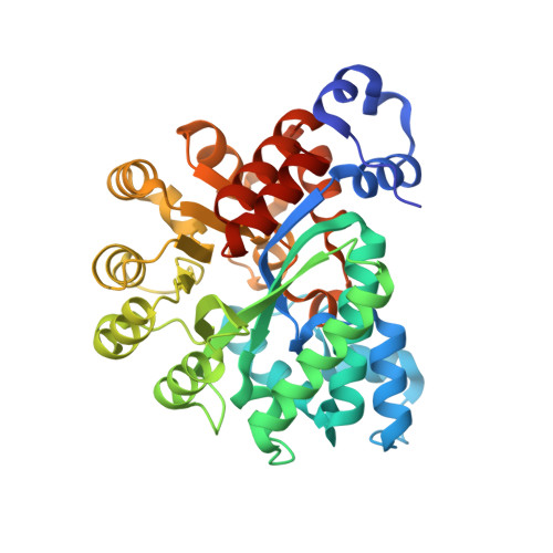
Plasmodium falciparum is a purine auxotroph requiring hypoxanthine as a key metabolic precursor. Erythrocyte adenine nucleotides are the source of the purine precursors, making adenosine deaminase (ADA) a key enzyme in the pathway of hypoxanthine formation. Methylthioadenosine (MTA) is a substrate for most malarial ADAs, but not for human ADA. The catalytic site specificity of malarial ADAs permits methylthiocoformycin (MT-coformycin) to act as a Plasmodium-specific transition state analogue with low affinity for human ADA [Tyler, P. C., Taylor, E. A., Frohlich, R. G. G., and Schramm, V. L. (2007) J. Am. Chem. Soc. 129, 6872-6879]. The structural basis for MTA and MT-coformycin specificity in malarial ADAs is the subject of speculation [Larson, E. T., et al. (2008) J. Mol. Biol. 381, 975-988]. Here, the crystal structure of ADA from Plasmodium vivax (PvADA) in a complex with MT-coformycin reveals an unprecedented binding geometry for 5'-methylthioribosyl groups in the malarial ADAs. Compared to malarial ADA complexes with adenosine or deoxycoformycin, 5'-methylthioribosyl groups are rotated 130 degrees . A hydrogen bonding network between Asp172 and the 3'-hydroxyl of MT-coformycin is essential for recognition of the 5'-methylthioribosyl group. Water occupies the 5'-hydroxyl binding site when MT-coformycin is bound. Mutagenesis of Asp172 destroys the substrate specificity for MTA and MT-coformycin. Kinetic, mutagenic, and structural analyses of PvADA and kinetic analysis of five other Plasmodium ADAs establish the unique structural basis for its specificity for MTA and MT-coformycin. Plasmodium gallinaceum ADA does not use MTA as a substrate, is not inhibited by MT-coformycin, and is missing Asp172. Treatment of P. falciparum cultures with coformycin or MT-coformycin in the presence of MTA is effective in inhibiting parasite growth.
Organizational Affiliation:
Department of Biochemistry, Albert Einstein College of Medicine, Yeshiva University, Bronx, New York 10461, USA.