3EB5

Structure of the cIAP2 RING domain


Experimental Data Snapshot

  • Method: X-RAY DIFFRACTION
  • Resolution: 2.00 Å
  • R-Value Free: 0.275 
  • R-Value Work: 0.213 
  • R-Value Observed: 0.219 

wwPDB Validation   3D Report Full Report


This is version 1.2 of the entry. See complete history


Literature

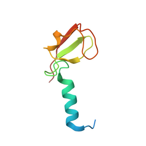
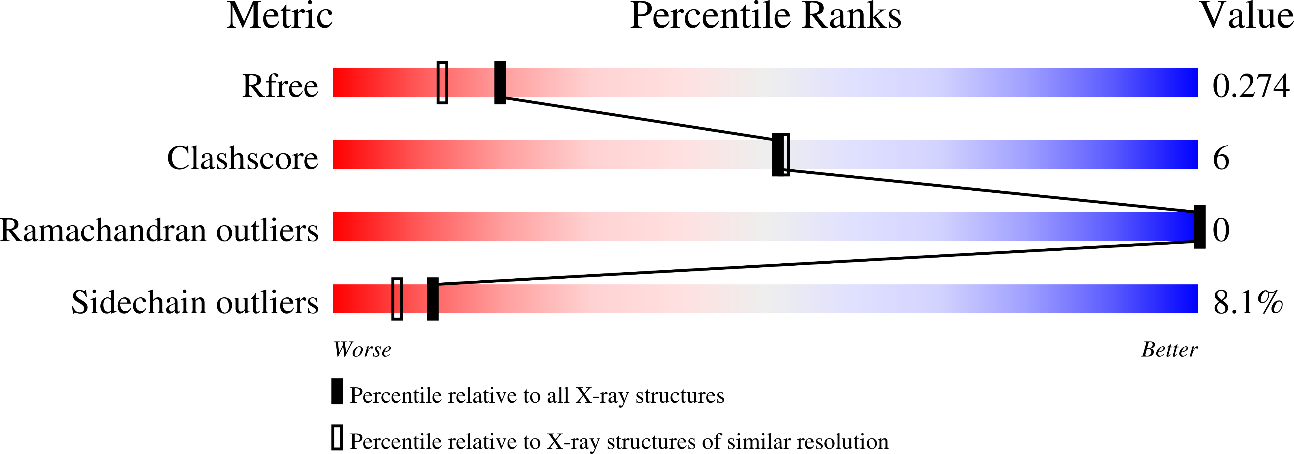
Structures of the cIAP2 RING domain reveal conformational changes associated with ubiquitin-conjugating enzyme (E2) recruitment.

Mace, P.D.Linke, K.Feltham, R.Schumacher, F.R.Smith, C.A.Vaux, D.L.Silke, J.Day, C.L.

(2008) J Biol Chem 283: 31633-31640

  • DOI: https://doi.org/10.1074/jbc.M804753200
  • Primary Citation of Related Structures:  
    3EB5, 3EB6

  • PubMed Abstract: 

    Inhibitor of apoptosis (IAP) proteins are key negative regulators of cell death that are highly expressed in many cancers. Cell death caused by antagonists that bind to IAP proteins is associated with their ubiquitylation and degradation. The RING domain at the C terminus of IAP proteins is pivotal. Here we report the crystal structures of the cIAP2 RING domain homodimer alone, and bound to the ubiquitin-conjugating (E2) enzyme UbcH5b. These structures show that small changes in the RING domain accompany E2 binding. By mutating residues at the E2-binding surface, we show that autoubiquitylation is required for regulation of IAP abundance. Dimer formation is also critical, and mutation of a single C-terminal residue abrogated dimer formation and E3 ligase activity was diminished. We further demonstrate that disruption of E2 binding, or dimerization, stabilizes IAP proteins against IAP antagonists in vivo.


  • Organizational Affiliation

    Biochemistry Department, University of Otago, Dunedin 9054, New Zealand.


Macromolecules
Find similar proteins by:  (by identity cutoff)  |  3D Structure
Entity ID: 1
MoleculeChains Sequence LengthOrganismDetailsImage
Baculoviral IAP repeat-containing protein 374Homo sapiensMutation(s): 0 
Gene Names: BIRC3API2IAP1MIHCRNF49
UniProt & NIH Common Fund Data Resources
Find proteins for Q13489 (Homo sapiens)
Explore Q13489 
Go to UniProtKB:  Q13489
PHAROS:  Q13489
GTEx:  ENSG00000023445 
Entity Groups  
Sequence Clusters30% Identity50% Identity70% Identity90% Identity95% Identity100% Identity
UniProt GroupQ13489
Sequence Annotations
Expand
  • Reference Sequence
Experimental Data & Validation

Experimental Data

  • Method: X-RAY DIFFRACTION
  • Resolution: 2.00 Å
  • R-Value Free: 0.275 
  • R-Value Work: 0.213 
  • R-Value Observed: 0.219 
  • Space Group: C 2 2 2
Unit Cell:
Length ( Å )Angle ( ˚ )
a = 34.869α = 90
b = 66.474β = 90
c = 77.465γ = 90
Software Package:
Software NamePurpose
SCALAdata scaling
REFMACrefinement
PDB_EXTRACTdata extraction
MOSFLMdata reduction

Structure Validation

View Full Validation Report


Entry History 

Deposition Data

Revision History  (Full details and data files)

  • Version 1.0: 2008-09-09
    Type: Initial release
  • Version 1.1: 2011-07-13
    Changes: Advisory, Version format compliance
  • Version 1.2: 2024-02-21
    Changes: Data collection, Database references, Derived calculations