3E46

Crystal structure of ubiquitin-conjugating enzyme E2-25kDa (Huntington interacting protein 2) M172A mutant


Experimental Data Snapshot

  • Method: X-RAY DIFFRACTION
  • Resolution: 1.86 Å
  • R-Value Free: 0.210 
  • R-Value Work: 0.172 
  • R-Value Observed: 0.174 

wwPDB Validation   3D Report Full Report


This is version 1.3 of the entry. See complete history


Literature

Structure of full-length ubiquitin-conjugating enzyme E2-25K (huntingtin-interacting protein 2).

Wilson, R.C.Hughes, R.C.Flatt, J.W.Meehan, E.J.Ng, J.D.Twigg, P.D.

(2009) Acta Crystallogr Sect F Struct Biol Cryst Commun 65: 440-444

  • DOI: https://doi.org/10.1107/S1744309109011117
  • Primary Citation of Related Structures:  
    3E46, 3F92

  • PubMed Abstract: 

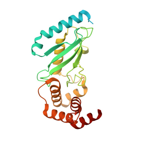
    The ubiquitin-conjugating enzyme E2-25K has been identified as a huntingtin (the key protein in Huntington's disease) interacting protein and has been shown to play a role in mediating the toxicity of Abeta, the principal protein involved in Alzheimer's disease pathogenesis. E2-25K is a dual-domain protein with an ubiquitin-associated (UBA) domain as well as a conserved ubiquitin-conjugating (UBC) domain which catalyzes the formation of a covalent bond between the C-terminal glycine of an ubiquitin molecule and the -amine of a lysine residue on the acceptor protein as part of the ubiquitin-proteasome pathway. The crystal structures of E2-25K M172A mutant protein at pH 6.5 and pH 8.5 were determined to 1.9 and 2.2 A resolution, respectively. Examination of the structures revealed domain-domain interactions between the UBC and UBA domains which have not previously been reported.


  • Organizational Affiliation

    University of Alabama in Huntsville, 35899, USA.


Macromolecules
Find similar proteins by:  (by identity cutoff)  |  3D Structure
Entity ID: 1
MoleculeChains Sequence LengthOrganismDetailsImage
Ubiquitin-conjugating enzyme E2-25 kDa253Homo sapiensMutation(s): 1 
Gene Names: UBE2KHIP2LIG
EC: 6.3.2.19
UniProt & NIH Common Fund Data Resources
Find proteins for P61086 (Homo sapiens)
Explore P61086 
Go to UniProtKB:  P61086
PHAROS:  P61086
GTEx:  ENSG00000078140 
Entity Groups  
Sequence Clusters30% Identity50% Identity70% Identity90% Identity95% Identity100% Identity
UniProt GroupP61086
Sequence Annotations
Expand
  • Reference Sequence
Small Molecules
Ligands 1 Unique
IDChains Name / Formula / InChI Key2D Diagram3D Interactions
CA
Query on CA

Download Ideal Coordinates CCD File 
B [auth A]CALCIUM ION
Ca
BHPQYMZQTOCNFJ-UHFFFAOYSA-N
Experimental Data & Validation

Experimental Data

  • Method: X-RAY DIFFRACTION
  • Resolution: 1.86 Å
  • R-Value Free: 0.210 
  • R-Value Work: 0.172 
  • R-Value Observed: 0.174 
  • Space Group: I 4
Unit Cell:
Length ( Å )Angle ( ˚ )
a = 134.49α = 90
b = 134.49β = 90
c = 38.404γ = 90
Software Package:
Software NamePurpose
MOLREPphasing
REFMACrefinement
PDB_EXTRACTdata extraction
HKL-2000data collection
DENZOdata reduction
SCALEPACKdata scaling

Structure Validation

View Full Validation Report


Entry History 

Deposition Data

Revision History  (Full details and data files)

  • Version 1.0: 2008-08-26
    Type: Initial release
  • Version 1.1: 2011-07-13
    Changes: Version format compliance
  • Version 1.2: 2021-10-20
    Changes: Database references, Derived calculations
  • Version 1.3: 2023-08-30
    Changes: Data collection, Refinement description