3DUZ

Crystal structure of the postfusion form of baculovirus fusion protein GP64


Experimental Data Snapshot

  • Method: X-RAY DIFFRACTION
  • Resolution: 2.95 Å
  • R-Value Free: 0.270 
  • R-Value Work: 0.224 
  • R-Value Observed: 0.226 

wwPDB Validation   3D Report Full Report


This is version 1.1 of the entry. See complete history


Literature

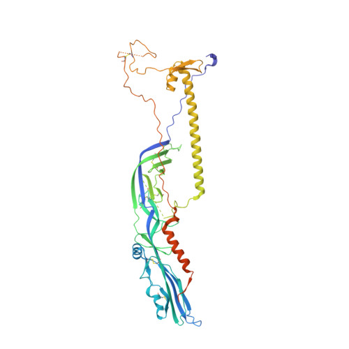
The postfusion structure of baculovirus gp64 supports a unified view of viral fusion machines.

Kadlec, J.Loureiro, S.Abrescia, N.G.Stuart, D.I.Jones, I.M.

(2008) Nat Struct Mol Biol 15: 1024-1030

  • DOI: https://doi.org/10.1038/nsmb.1484
  • Primary Citation of Related Structures:  
    3DUZ

  • PubMed Abstract: 

    Viral fusion proteins mediate the merger of host and viral membranes during cell entry for all enveloped viruses. Baculovirus glycoprotein gp64 (gp64) is unusual in promoting entry into both insect and mammalian cells and is distinct from established class I and class II fusion proteins. We report the crystal structure of its postfusion form, which explains a number of gp64's biological properties including its cellular promiscuity, identifies the fusion peptides and shows it to be the third representative of a new class (III) of fusion proteins with unexpected structural homology with vesicular stomatitis virus G and herpes simplex virus type 1 gB proteins. We show that domains of class III proteins have counterparts in both class I and II proteins, suggesting that all these viral fusion machines are structurally more related than previously thought.


  • Organizational Affiliation

    Division of Structural Biology, Wellcome Trust Centre for Human Genetics, University of Oxford, Roosevelt Drive, Oxford OX3 7BN, UK.


Macromolecules
Find similar proteins by:  (by identity cutoff)  |  3D Structure
Entity ID: 1
MoleculeChains Sequence LengthOrganismDetailsImage
Major envelope glycoprotein487Autographa californica nucleopolyhedrovirusMutation(s): 0 
Gene Names: GP67P67
Membrane Entity: Yes 
UniProt
Find proteins for P17501 (Autographa californica nuclear polyhedrosis virus)
Explore P17501 
Go to UniProtKB:  P17501
Entity Groups  
Sequence Clusters30% Identity50% Identity70% Identity90% Identity95% Identity100% Identity
UniProt GroupP17501
Sequence Annotations
Expand
  • Reference Sequence
Experimental Data & Validation

Experimental Data

  • Method: X-RAY DIFFRACTION
  • Resolution: 2.95 Å
  • R-Value Free: 0.270 
  • R-Value Work: 0.224 
  • R-Value Observed: 0.226 
  • Space Group: H 3 2
Unit Cell:
Length ( Å )Angle ( ˚ )
a = 87.47α = 90
b = 87.47β = 90
c = 431.59γ = 120
Software Package:
Software NamePurpose
REFMACrefinement
DNAdata collection
XDSdata reduction
XSCALEdata scaling
SHARPphasing

Structure Validation

View Full Validation Report


Entry History 

Deposition Data

Revision History  (Full details and data files)

  • Version 1.0: 2008-09-16
    Type: Initial release
  • Version 1.1: 2011-07-13
    Changes: Version format compliance