3DIT

Crystal structure of MAD MH2 domain


Experimental Data Snapshot

  • Method: X-RAY DIFFRACTION
  • Resolution: 3.20 Å
  • R-Value Free: 0.310 
  • R-Value Work: 0.244 

wwPDB Validation   3D Report Full Report


This is version 1.2 of the entry. See complete history


Literature

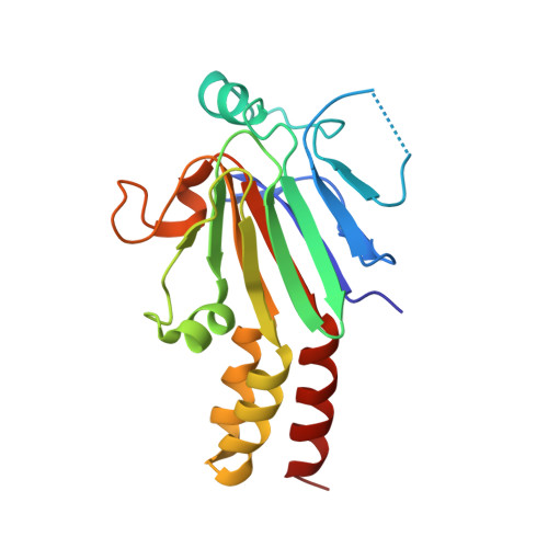
Structure of Drosophila Mad MH2 domain.

Hao, R.Chen, L.Wu, J.W.Wang, Z.X.

(2008) Acta Crystallogr Sect F Struct Biol Cryst Commun 64: 986-990

  • DOI: https://doi.org/10.1107/S1744309108033034
  • Primary Citation of Related Structures:  
    3DIT

  • PubMed Abstract: 

    In Drosophila, decapentaplegic (Dpp), a member of the TGF-beta superfamily, plays a pivotal role in control of proliferation, global patterning and induction of specific cell fates. Together with Medea, mother against Dpp (Mad), the founding member of the Smad family, specifically transduces the Dpp signal from the plasma membrane to the nucleus. Here, the crystal structure of the MH2 domain of Mad, which closely matches those of other Smad MH2 domains, is reported at 3.2 A resolution. The conservation of Smad protein structures is consistent with their evolutionary conserved and significant function. Furthermore, sequence alignment revealed that most of the variant amino acids in Smad proteins specific to the BMP pathway (Smad1, Smad5 and Mad) were clustered at the surface. In particular, Ser296 and Asp297 of Mad introduced a negative patch into the positive surface observed in the surface electrostatic potential of Smad1 MH2.


  • Organizational Affiliation

    Institute of Biophysics and Graduate University, Chinese Academy of Sciences, Beijing 100101, People's Republic of China.


Macromolecules
Find similar proteins by:  (by identity cutoff)  |  3D Structure
Entity ID: 1
MoleculeChains Sequence LengthOrganismDetailsImage
Protein mothers against dpp
A, B, C
188Drosophila melanogasterMutation(s): 0 
Gene Names: MadCG12399
UniProt
Find proteins for P42003 (Drosophila melanogaster)
Explore P42003 
Go to UniProtKB:  P42003
Entity Groups  
Sequence Clusters30% Identity50% Identity70% Identity90% Identity95% Identity100% Identity
UniProt GroupP42003
Sequence Annotations
Expand
  • Reference Sequence
Experimental Data & Validation

Experimental Data

  • Method: X-RAY DIFFRACTION
  • Resolution: 3.20 Å
  • R-Value Free: 0.310 
  • R-Value Work: 0.244 
  • Space Group: P 32
Unit Cell:
Length ( Å )Angle ( ˚ )
a = 95.864α = 90
b = 95.864β = 90
c = 66.726γ = 120
Software Package:
Software NamePurpose
CNSrefinement
PDB_EXTRACTdata extraction
HKL-2000data collection
HKL-2000data reduction
HKL-2000data scaling
PHASERphasing

Structure Validation

View Full Validation Report


Entry History 

Deposition Data

Revision History  (Full details and data files)

  • Version 1.0: 2009-05-12
    Type: Initial release
  • Version 1.1: 2011-07-13
    Changes: Version format compliance
  • Version 1.2: 2023-11-01
    Changes: Data collection, Database references, Refinement description